Download

1 / 9
0 likes | 0 Views
Diabetic patients frequently suffer from non-alcoholic steatohepatitis. The current study aimed to investigate the role of curcumin and the response of hepatic stellate cells in streptozotocin (STZ)-induced hepatic damage. Sixty male rats were divided into three groups. The normal control injected with a citrate buffer vehicle and the diabetic control group which was injected intraperitoneally (IP) with a single-dose of streptozotocin (50mg/kg body weight) and a diabetic group was treated with an oral dose of curcumin at 80 mg/kg body weight daily for 60 days. Curcumin effectively counteracts

E N D
Tissue and Cell 48 (2016) 81–88 Contents lists available at ScienceDirect Tissue and Cell jour nal ho mepage: www.elsevier.com/locate/tice The the role of curcumin in streptozotocin-induced hepatic damage and trans-differentiation of hepatic stellate cells Hesham N. Mustafa∗ Anatomy Department, Faculty of Medicine, King Abdulaziz University, Jeddah, Saudi Arabia a r t i c l e i n f o a b s t r a c t Article Received Received Accepted Available history: Diabetic tigate hepatic citrate single-dose dose stress-mediated SMA) curcumin is it cells cell curcumin’s patients frequently suffer from non-alcoholic steatohepatitis. The current study aimed to inves- 5 December 2015 the role ofcurcumin and the response of hepatic stellate cells in streptozotocin (STZ)-induced inrevised form 19January 2016 damage. Sixty male rats were divided into three groups. The normal control injected with a 7February 2016 buffer vehicle and the diabetic control group which was injected intraperitoneally (IP) with a online 9February 2016 of streptozotocin (50 mg/kg body weight) and a diabetic group was treated with an oral of curcumin at 80 mg/kg body weight daily for 60 days. Curcumin effectively counteracts oxidative Keywords: Curcumin Hepatic Streptozotocin Lipid Trans-differentiation hepatic damage and improves biochemical parameters. Alpha-smooth muscle actin (?- was significantly reduced, and insulin antibodies showed strong positive immunoreactivity with stellate cells administration. These results optimistically demonstrate the potential use of curcumin, which attributed to its antiradical/antioxidant activities and its potential ?-cell regenerative properties. Also, peroxidation has the capability to encourage the trans-differentiation of hepatic stellate cells into insulin-producing for a period of time. In addition, as it is an anti-fibrotic mediator that inhibits hepatic stellate activation and the transition to myofibroblast-like cells, this suggests the possibility ofconsidering novel therapeutic effects in reducing hepatic dysfunction in diabetic patients. © 2016 Elsevier Ltd.All rights reserved. 1. Introduction leads hepatic RAGEs. Oxidative of tion Ponmurugan, The liver tion antioxidant, curcuminoid eraceae The tion C, 2010). lipidemia signal pathways This stellate liver in dysfunction. toan increased proliferation of HSCs, which isnoticed during fibrogenesis that isaccompanied by the up-regulation of Diabetic recognized with cirrhosis, 2007). Streptozotocin of the dependent Alpha-smooth recognition (HSCs) In to (AGEs). consequently patients frequently suffer from ahepatic impairment as non-alcoholic steatohepatitis which isassociated stress plays acrucial role inthe chronic complications severe complications such as deposition of glycogen, steatosis, the diabetic liver, where itisassociated with an overproduc- fibrosis and occasionally hepatic cancer (Bugianesi et al., of oxygen free radicals and lipid peroxidation (Saravanan and 2011). (STZ) is anN-nitroso-N-methylurea derivative use of herbal medicine indefending against STZ-induced 2-deoxy-d-glucose which isadiabetogenic agent that damages damage looks promising. Curcumin has been gaining atten- islet ?-cells inthe pancreas selectively toproduce insulin- because of its health benefits, such as its anti-inflammatory, diabetes mellitus (IDDM) (Yang et al., 2010). and immune modulatory effects. Curcumin isthe chief muscle actin (?-SMA) isan indicator for the of Longa, which is amember of the Zingib- Curcuma of myofibroblast-like cells plus hepatic stellate cells family (Kumar etal., 2015). which are also known as Itocells (Clement et al., 2010). polyphenol curcumin improves diabetes-induced dysfunc- diabetes, the glucose accessibility isincreased and this leads by decreasing the level of glucose, inhibiting protein-kinase an accelerated formation of advanced glycation end products and lowering superoxide production (Rungseesantivanon et al., AGEs interact with the receptor for AGEs (RAGEs) and Itreverses insulin resistance, hyperglycemia, and hyper- increase oxidative stress and cellular growth. This by inhibiting the pro-inflammatory transcription factors, transducers and stimulating anti-inflammatory signaling (Tang and Chen, 2010). study aimed toinvestigate the probable influence of hepatic cells inthe protective capability of curcumin inSTZ-induced ∗Corresponding Anatomy Saudi E-mail author at: King Abdulaziz University, Faculty of Medicine, damage. Itwas also intended toprovide an important support Department, Ground Floor Building No. 8, P.O. Box: 80205, Jeddah 21589, understanding the mechanism ofcurcumin treatment for hepatic Arabia. Tel.: +966 566 764 762. hesham977@hotmail.com address: http://dx.doi.org/10.1016/j.tice.2016.02.003 0040-8166/© 2016 Elsevier Ltd. All rights reserved.
82 H.N. Mustafa /Tissue and Cell 48(2016) 81–88 2. Materials and methods 2.8. Serum parameters ALT, AST, and ALP, which were increased following hepatocyte 2.1. Ethical approval injury, manuals lin, bilirubin responding (Cairo, were assessed according tothe protocol detailed inthe This study was conducted after receiving the approval of the of the diagnostic kits (Randox Laboratories Ltd., Crum- Medical Abdulaziz Research Ethics Committee, Faculty of Medicine, King County Antrim, UK). Serum albumin, total protein, and total University, Jeddah, Saudi Arabia. were determined by spectrophotometer using the cor- colorimetric kits supplied by Bio Diagnostic Company Egypt) (Lee et al., 2012). 2.2. Chemicals and Reagents Streptozotocin Sigma–Aldrich transferase phosphatase Ltd. and nostic (STZ) and curcumin were purchased from 2.9. Histological examination Chemicals (St. Louis, MO, USA). Alanine amino- (ALT), aspartate aminotransferase (AST), and alkaline Livers were washed with a phosphate buffer solution and then (ALP) kits were purchased from Randox Laboratories fixed through ded inthickness eosin Acid-Schiff at BX53 Japan). in10% neutral buffered formalin. Tissues were dehydrated (Crumlin, County Antrim, UK). Serum albumin, total protein, agraded series ofalcohol, cleared inxylene, and embed- total bilirubin colorimetric kits were supplied bythe Bio Diag- inparaffin wax. Tissues were then cut into sections of 3–5?m Company (Cairo, Egypt). using amicrotome and stained with hematoxylin and (H&E) for histopathological evaluation and with periodic (PAS) for observation of glycogen. For each specimen, 2.3. Animals least three tofive slides were examined using an Olympus Sixty male adult albino Wistar rats, weighing 190 ±20 g, that microscope equipped with aDP73 camera (Olympus, Tokyo, were housed 40–70% standard The the alaboratory. used were obtained from the animal house. Animals were ina(24◦C± 3◦C) temperature-controlled room with humidity and 12/12 hlight/dark cycle. Rats were fed a 2.10. Histopathological evaluation diet and tap water throughout the experiment. adlibitum experimental procedures were performed inaccordance with The sections were analyzed for hydropic swelling, parenchy- international guidelines for the care and the use of animals in matous vacuole, sinusoids presented (Guven lent represented mals 2015). degeneration, microvesicular vacuole, macrovesicular focal necrosis, inflammatory infiltrations, fibrosis, and hyperemia. At the end of the analyses, the findings were 2.4. Induction of diabetes inatable which showed the degree of degeneration et al., 2006). Score levels of 0, +1, +2, +3 were equiva- Fasted rats (12 h)received asingle intraperitoneal (IP) injection tono, mild, moderate, and severe, respectively. The scores of citrate cose the obtaining tic Blood freshly prepared STZ (50 mg/kg body weight) dissolved in0.1 M values obtained from the tissue sections of six ani- buffer (pH 4.5). STZ-injected animals were given a5%glu- from each group with five fields/section (Mustafa et al., solution for 24htoovercome drug-induced hypoglycemia. On third day after STZ injection, glucose levels were estimated by blood samples from the cut tipof the tail using Diagnos- 2.11. Immunohistochemical examination Accu-Chek test strips (Roche Diagnostics, Mannheim, Germany). glucose levels of 250 mg/dl or more were considered diabetic. The standard peroxidase immunohistochemistry technique was applied de-waxed gen Microwave-assisted Slides antibody Carpinteria, cytoplasmic) of similarly mouse with were peroxidase Sections and substrate substrate were sections control the microscopy, Absence the (+) toslides of paraffin-embedded tissue. Sections were 2.5. Experimental design inxylene, rehydrated, and pretreated with 3%of hydro- peroxide solution toblock endogenous peroxidase activity. The animals were distributed into 3groups (20/group). Group antigen retrieval was performed for 20min. 1(normal Group (50 STZ injection, body the retro-orbital ysis. processed control) was injected IPwith acitrate buffer vehicle. were then incubated overnight at4◦Cwith the primary 2(diabetic control) received asingle IPinjection of STZ against ?-SMA (amouse monoclonal antibody [Dako, mg/kg body weight). Group 3received asingle IP injection of California, USA] with adilution of 1:50; cellular site was (50 mg/kg body weight) and, on the third day after the STZ as amarker ofactivated HSCs with varying degrees curcumin was given orally with a dosage of 80mg/kg intensity insmooth muscles and myofibroblasts. They were weight and continued daily for 60 days (Zhang etal., 2013). At incubated with the primary antibody against insulin (a end of the experiment, blood samples were collected from the monoclonal antibody [Dako, Carpinteria, California, USA] sinus in heparinized capillary tubes for serum anal- adilution of 1:100; cellular site was cytoplasmic). The sections Animals from all groups were sacrificed, and the livers were incubated with biotinylated IgG and then with streptavidin- for histological studies. conjugate (Zymed Corp, San Francisco, CA, USA). were then washed with phosphate-buffered saline (PBS) 2.6. Plasma glucose estimation incubated with 3, 3?-diaminobenzidine tetrachloride (DAB) chromogen solution (1drop of DAB chromogen/1 mL of Plasma glucose was measured using an enzymatic colorimet- buffer) for 5min todetect immunoreactivity. All sections ric Ltd., method with commercially available kits (Randox Laboratories, counter-stained with Mayer’s hematoxylin. Negative control Antrim, UK). were prepared by omitting the primary antibody. Positive standard laboratory slides were used for all stains toprove success of the technique. All slides were examined under light 2.7. Plasma insulin estimation and the presence of labeled cells was documented. Plasma immunosorbent Co., insulin was determined using an insulin enzyme-linked of staining was recognized as anegative result (−), while assay (ELISA) kit (code no. AKRIN-010T; Shibayagi presence of brown staining was recognized as positive result Ltd., Gunma, Japan). (Mustafa et al., 2015).
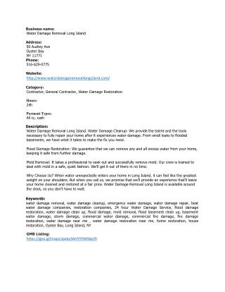
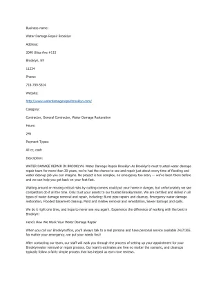
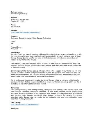
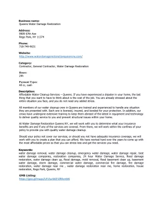
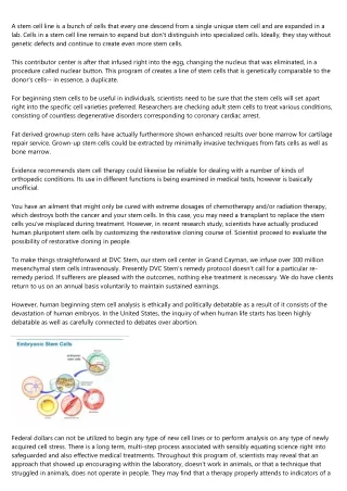
83 H.N. Mustafa /Tissue and Cell 48(2016) 81–88 Table Body 1 and liver weight of different groups. Normal control (n=20) STZ-diabetic control (n=20) STZ-diabetes +curcumin (n=20) Liver weight (g) 11.14 ±1.54 9.91 P* ±1.04 10.83 *P **P 180.47 *P **P ±0.85 <0.01 >0.05 <0.05 Body weight (g) 187.65 ±6.11 163.94 *P ±4.9 3.89 ± <0.001 <0.001 <0.001 Values *P: are means ±SD(n=20 each group). ANOVA followed byTukey’s post hoc test. compared tonormal control. compared todiabetic control. **P: Fig. 1.Body and liver weight of different groups. The mean isgiven incolumns, and error bars represent the standard deviation (SD). albumin curcumin eters the and Fig. and total protein in the diabetic control group, while 2.12. Quantitative morphometric measurements treatment showed a significant increase inthese param- non-overlapping criminately Image-Pro MD, nih.gov/ij/)with Japan). the 2015). Ten fields for each animal were selected indis- compared tothose of the diabetic control group. Conversely, and analyzed. The measurements were done by using total bilirubin was increased inthe diabetic control group Plus v6.0 software (Media Cybernetics, Silver Spring, decreased significantly with curcumin treatment (Table 2and USA) and the NIH ImageJ (v1.50) program (http://rsb.info. 3). anOlympus BX53 microscope (Olympus, Tokyo, The area percentages of ?-SMA immunopositive cells and 3.3. Histopathological findings insulin-positive cells number were evaluated (Hussein et al., The normal control group showed normal cytoarchitecture. The diabetic loss were PAS-stained positive more of (Fig. were control group showed parenchymatous degeneration and 2.13. Statistical analysis of architecture. With curcumin treatment, hepatic changes reduced (Table 3). Quantitative parameters one-way test. 23. data were expressed as the mean SD of different sections of the normal control group showed PAS- ± for the treated groups. The data were analyzed using red granules inthe cytoplasm of the hepatocytes and analysis of variance (ANOVA) followed by Tukey’s post hoc granules around the central vein (Fig. 4A), while those The statistical analysis was performed using IBM SPSS version the diabetic control group showed few PAS-positive granules The values were considered significant when p<0.05. 4B). With curcumin treatment, mild PAS-positive granules observed (Fig. 4Cand Table 3). 3. Results 3.4. Immunohistochemistry-stained sections 3.1. Effect on liver and body weight Inthe normal control group, ?-SMA staining was observed only At the end of 60 days, curcumin treated specimens showed a in branches, group the soidal mild age control reduced Insulin-immunostained (Fig. mal showed around cells and the media of the blood vessels (portal veins, hepatic artery significant the increase inthe body and liver weight as compared to and central veins) (Fig. 5A). While the diabetic control diabetic control (Table 1and Fig. 1). showed positive ?-SMA immunoreactivity inthe media of portal vessels and the periportal area and along the perisinu- spaces (Fig. 5Band C). Inthe curcumin treatment group, 3.2. Biochemical parameters immunoreactivity was observed (Fig. 5D). The area percent- of ?-SMA positive cells was significantly higher in the diabetic The mean glucose level of the diabetic control group was signif- group, while inthe curcumin treatment group itwas icantly improvement level curcumin level Plasma control while significantly. increased while curcumin treatment showed asignificant significantly (Table 4and Fig. 7). in glucose level (Table 2and Fig. 2). The mean insulin sections for both the normal control of the diabetic control group was significantly decreased while 6A) and diabetic control (Fig. 6B) groups showed no ormini- treatment showed asignificant improvement ininsulin insulin immune positive cells. The curcumin treatment group (Table 2and Fig. 2). many insulin positive immune cells inthe portal area ALT, AST, and ALP levels were elevated inthe diabetic the bile ductule (Fig. 6C). The number of insulin positive group incomparison tothose of the normal control group, was significantly higher inthe curcumin treated group (Table 4 with curcumin treatment all of these parameters are reduced Fig. 7). Furthermore, there was asignificant decrease inthe
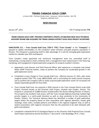
84 H.N. Mustafa /Tissue and Cell 48(2016) 81–88 Fig. 2. Plasma glucose and insulin of different groups. The mean isgiven incolumns, and error bars represent the standard deviation (SD). Table Biochemical 2 parameters of the different groups. Parameters Normal control STZ-diabetic control STZ-diabetes + curcumin Plasma glucose (mg/dl) 87.14 ±4.28 280.12 *P ±57.24 160.12 *P **P 4.06 *P **P 76.8 *P **P 204.68 *P **P 79.5 *P **P 3.81 *P **P 4.62 *P **P 0.36 *P *P ±63.27 <0.001 <0.001 <0.001 Plasma insulin (mIU/L) 5.17 ±0.73 0.54 *P ±0.11 ±0.45 <0.001 <0.001 < 0.001 ALT (U/l) 65.4 ±1.2 119.6 *P ±2.30 ±4.60 <0.001 <0.001 <0.001 AST (U/l) 139.13 ±3.82 321.35 *P ±2.80 ±1.96 <0.001 <0.001 <0.001 ALP (U/l) 68.10 ±2.30 85.2 *P 4.60 ±8.10 ± <0.001 <0.001 <0.01 Albumin (g/dl) 4.14 ±0.60 1.3 *P ±0.87 ±0.40 <0.001 >0.05 <0.001 Total protein (g/dL) 5.94 ±0.89 2.03 *P ±0.78 ±0.13 <0.001 <0.001 <0.001 Total bilirubin (mg/dL) 0.24 ±0.20 0.78 *P ±0.07 ±0.06 <0.001 <0.05 <0.001 Values *P: ALT, units are means ±SD(n=20 each group). ANOVA followed byTukey’s post hoc test. compared tonormal control. compared todiabetic control. **P: alanine aminotransferase; AST, aspartate aminotransferase; ALP, alkaline phosphatase; mg/dl, milligrams per deciliter; mIU/L, milli-international units per liter; U/l, per liter; g/dl, grams per deciliter. 4. Discussion mainly consensus cumin In gestion near are stores and radicals antioxidants some tissue is In undergo myofibroblast-like extracellular (She areduction cell improvement Curcumin tion RAGE tumor necrosis factor (TNF-?), interleukin-1 (IL-1), and interferon (IFN-c) (Sugano et al., 2006). Meanwhile, cur- Inthe current study, the diabetic control group revealed areduc- treatment alleviates the hepatocellular injuries. tion catabolism 2010). this tural processes The insulin 2013).Improved cumin 2007), itis regeneration tothe (Montanya Hepatocellular (ROS) tocytes Rahmat, ucts, fibrosis inflammatory inbody weight, which isattributed tothe muscle destruction or the present study, the diabetic control group revealed acon- of structural protein and fat (Salahuddin and Jalalpure, of the portal triad with inflammation and notable fibrosis Curcumin administration improves the body weight, and the central vein. This isattributed totissue degradations that indicates the effect of curcumin on the degradation of struc- caused by depletion of the endogenous antioxidant enzyme protein, control of muscle wasting, and increase of metabolic as superoxide dismutase (SOD) and catalase (CAT) (Chang inthe body. Chuang, 2010)and promotion of de novo generation of free current data showed a significant decrease inthe plasma (Maritim et al., 2003).Consequently, the higher level of levels, which coincides with other findings (Abdel Aziz etal., that exists incurcumin could boost the quenching of insulin level supports the hypothesis that cur- free radicals inside hepatocytes, which defend the hepatic causes ?-cells neogenesis and protection (Meghana et al., against oxidative stress damage (Afrin et al., 2015),and this and this was evidently proven by this study. Furthermore, supported by the impairment, current findings. hepatic proposed that controlling hyperglycemia encourages further liver stellate cells (Ito cells) of ?-cells (Guz et al., 2001).This may be attributed trans-differentiation from lipid storing pericytes to suggestion that dying ?-cells are regenerated continuously cells that lead toan increased collagen and et al., 2000). matrix that isthe crucial event inhepatic fibrogenesis injury iscredited toreactive oxygen species etal., 2005). Curcumin treatment inthe current study caused and lipid peroxidation that causes direct damage tohepa- of?-SMA positive cells, which is the marker of Ito by disrupting the membranes, protein, and DNA (Asmah activation and inturn, indicates inhibition of Itocells and the 2015).The injured hepatocytes release aldehyde end prod- of hepatic fibrosis (Erenoglu et al., 2011). which causes further hepatocellular damage and emergence of suppresses the AGEs mediated by the stimula- (Novo and Parola, 2012). Also, lipid peroxidation causes an of RAGEs gene expression. This leads toinhibition of the reaction that involves the occurrence of cytokines, activated pathways which, inturn, prevents oxidative
85 H.N. Mustafa /Tissue and Cell 48(2016) 81–88 Fig. 3. Biochemical parameters of different groups. The mean is given incolumns, and error bars represent the standard deviation (SD). Table Histopathological 3 Inhyperglycemic current stellate tion maintain trans-differentiation diabetic fact primitive the might pancreatic In existence the tion hyperglycemic support attributed curcumin were maintain cells These of because regenerative differentiation for that conditions, the diabetic control group in the findings of the liver. study showed minimal insulin immune reactions of hepatic cells. This indicates that diabetes encourages the transi- Normal control STZ-diabetic control STZ- diabetes curcumin + of hepatic stellate cells toinsulin-producing cells but itcannot their production. Furthermore, itwas found that this occurs early and lasts for two weeks inthe Hydropic Parenchymatous Microvesicular Macrovesicular Focal Inflammatory Fibrosis Sinusoids PAS swelling 0 0 0 0 0 0 0 0 +2 +2 +0.5 +0.5 +2 +1 +1 +2 +1 +1 +1 +1 +1 +0.5 +1 +0.5 +1 +2 degeneration liver (Kim etal., 2007).This transition is attributed tothe vacuole that both the liver and the pancreas originate from the upper vacuole foregut endoderm appendages (Lammert etal., 2003)and necrosis late separation of the pancreas and liver during organogenesis infiltrations leave pluripotent cells, which are able togive rise toboth hyperemia and hepatic lineage (Meivar-Levy and Ferber, 2003). +3 supporting this hypothesis, aprevious study has discussed the N=20; Scale: No (0), mild (+1), moderate (+2), severe (+3). of extra-pancreatic insulin-producing cells inthe liver in hyperglycemic condition. However, the number and produc- of these insulin-producing cells are not enough toimprove the condition (Kojima et al., 2004).The current results stress, mark associated over, lipid (Stefanska, Inthe myofibroblast-like between and cause factor growth Kupffer inflammation, and hepatic stellate cell activation, ahall- the hypothesis that the hypoglycemic effect of curcumin is of non-alcoholic steatohepatitis and hepatic fibrogenesis topreserving functional insulin-producing cells. Inthe with type 2diabetes mellitus (Stefanska, 2012).More- group inthe current study, insulin immune positive cells AGE-encouraged production of reactive oxygen species and increased which supports the suggestion that curcumin may peroxides was attenuated after treatment with curcumin insulin production by trans-differentiated hepatic stellate 2012). for aperiod after STZ injection. current findings, the trans-differentiation of Ito cells into results optimistically demonstrate the potential use cells isattributed tointercellular connections curcumin for the treatment ofSTZ-induced liver damage Itocells and activated Kupffer cells, damaged hepatocytes, of its antiradical/antioxidant activities, potential ?-cell inflammatory cytokines (Hellerbrand, 2013).Other factors that properties, and capability toencourage the trans- this trans-differentiation include the platelet-derived growth ofhepatic stellate cells into insulin-producing cells (PDGF), epidermal growth factor (EGF), and transforming aperiod of time. In addition, as itis an anti-fibrotic mediator factor ?1(TGF-?1) that are released after activation of inhibits hepatic stellate cell activation and the transition to cells (Nosseir etal., 2012).
86 H.N. Mustafa /Tissue and Cell 48(2016) 81–88 Fig. (C) 4. (A) Normal control group showed PAS-positive reaction (distribution of liver glycogen) (arrow); (B) diabetic control group showed fewPAS-positive reaction (arrow); curcumin treatment group showed mild PAS-positive reaction (arrow) (PAS, scale bar 20 ?m). Fig. portal treatment 5. (A) Normal control group showed minimal immunostaining (arrow); (B) diabetic control group showed positive immunostaining (activated HSCs) inthe media of the blood vessels and scattered inthe periportal area (arrow); (C) the previous group with positive immunostained cells inthe perisinusoidal spaces (arrow); (D) curcumin group showed mild immunostaining (arrow) (?-SMA, scale bar 20 ?m).
87 H.N. Mustafa /Tissue and Cell 48(2016) 81–88 Table Mean 4 ±SDof the area percentage of ?-smooth muscle actin positive cells and the number of insulin-positive cells. Normal control STZ-diabetic control STZ-diabetes +curcumin Area %of ?-SMA 0.43 ±0.28 10.20 *P ±0.62 1.06 *P **P 5.10 *P **P 0.87 ± <0.001 <0.01 <0.001 Number of insulin positive cells 0.00 0.28 *P ±0.01 3.50 ± >0.05 <0.001 <0.001 Values *P: are means ±SD(n=20 each group). ANOVA followed byTukey’s post hoc test. compared tonormal control. compared todiabetic control. **P: Fig. showed 6.(A) Normal control group showed anegative immune reaction; (B) diabetic control group showed aminimal immune reaction; (C) the curcumin treatment group strong positive immunoreactivity (insulin-producing cells) (arrows) (insulin immune reaction, scale bar 20 ?m). Fig. 7. Area %of ?-SMA and number of insulin positive cells. The mean isgiven incolumns, and error bars represent the standard deviation (SD). Conflicts of interest myofibroblast-like consider dysfunction cells, consequently the way is now open to curcumin’s novel therapeutic effects inreducing hepatic indiabetic patients. None. 5. Conclusion References The current data suggest that curcumin affects hepatic stel- Abdel Aziz, H.H., on term M.T., El-Asmar, M.F., Rezq, A.M., Mahfouz, S.M., Wassef, M.A., Fouad, Ahmed, H.H., Taha, F.M., 2013. The effect of a novel curcumin derivative late steatohepatitis cell activation, which represents akey event of non-alcoholic pancreatic islet regeneration inexperimental type-1 diabetes inrats (long and hepatic fibrogenesis associated with diabetes. study). Diabetol. Metab. Syndr. 5, 75.
88 H.N. Mustafa /Tissue and Cell 48(2016) 81–88 Arumugam, Karuppagounder, Nomoto, streptozotocin-induced reticulum 279–289. Asmah kidney Bugianesi, hepatocellular 175–180. Chang, type Transl. Clement, F., muscle 52,635–643. Erenoglu, Protective rats.Balkan Guven, Effects Acta Guz, intra-islet 142,4956–4968. Hellerbrand, 465,775–778. Hussein, combination testicular 325–334. Kim, Streptozotocin-induced activation regeneration. Kojima, Extrapancreatic Natl. Kumar, Curcumin Lammert, pancreas Lee, Liver Maritim, stress, Afrin, R., S., Soetikno, V., Thandavarayan, R.A., Pitchaimani, V., Meghana, streptozotocin-induced prophylactic Meivar-Levy, liver-to-pancreas 460–466. Montanya, beta-cell beta-cell Mustafa, coenzyme inrats: 47, Nosseir, protective tetrachloride-induced 398–411. Novo, healing Rungseesantivanon, Curcumin dysfunction PKCinhibition. Salahuddin, Cucumis Ethnopharmacol. Saravanan, oxidative 100–106. She, regulation Stefanska, mellitus 2209–2211. Sugano, Iino,N., High-fat induces diabetic Tang, stellate glucokinase. Yang, by streptozotocin 1415–1422. Zhang, review. K., Sanjeev, G., Ramesh, B., 2007. Curcumin prevents V., Sreedhar, R., Harima, M., Suzuki, H., Miyashita, S., islet damage by scavenging free radicals: a M., Suzuki, K., Watanabe, K., 2015. Curcumin ameliorates and protective role. Eur. J.Pharmacol. 577, 183–191. liver damage through modulation of endoplasmic I.,Ferber, S., 2003. New organs from our own tissues: stress-mediated apoptosis indiabetic rats. Free Radic. Res. 49, transdifferentiation. Trends Endocrinol. Metab. 14, Rahmat, A.A., 2015. Effect of pomegranate on histopathology of liver and E., Nacher, V., Biarnés, M., Soler, J., 2000. Linear correlation between on generated oxidative stress diabetic induced rats. J.Cytol. Histol. 6, 2. mass and body weight throughout the lifespan inLewis rats: role of E., Vanni, E., Marchesini, G., 2007. NASH and the risk of cirrhosis and hyperplasia and hypertrophy. Diabetes 49, 1341–1346. carcinoma intype 2diabetes. H., ElAwdan, S., Curr. Diab. Rep. 7, Hegazy, G., Abdel Jaleel, G., 2015. Prophylactic role of Q10 and Cynara scolymus Lon doxorubicin-induced toxicity Y.C., Chuang, L.M., 2010. The role of oxidative stress inthe pathogenesis of Biochemical and immunohistochemical study. Indian J.Pharmacol. 2diabetes: from molecular mechanism toclinical implication. Am. J. 649. Res. 2, 316–331. D.A., Gawish, S.A., Moustafa, A.M., Firgany, A.E.-D.L., 2012. The possible S., Pascarella, S., Conzelmann, S., Gonelle-Gispert, C., Guilloux, K., Negro, effect of green tea versus interferon-?on carbon 2010. The hepatitis Cvirus core protein indirectly induces alpha-smooth hepatic fibrosis inalbino rat. Egypt. J.Histol. 35, actin expression inhepatic stellate cells via interleukin-8. J.Hepatol. E., Parola, M., 2012. The role of redox mechanisms inhepatic chronic wound C., Kanter, M., Aksu, B., Sagiroglu, T., Ayvaz, S., Aktas, C., Erboga, M., 2011. and fibrogenesis. Fibrogenesis Tissue Repair 5, S4. effect of curcumin onliver damage induced by biliary obstruction in S., Thenchaisri, N., Ruangvejvorachai, P., Patumraj, S., 2010. Med. J.28, 352–357. supplementation could improve diabetes-induced endothelial A., Yavuz, O., Cam, M., Ercan, F., Bukan, N., Comunoglu, C., Gokce, F., 2006. associated with decreased vascular superoxide production and of melatonin onstreptozotocin-induced diabetic liver injury inrats. BMC Complement. Altern. Med. 10, 57. Histochem. 108, 85–93. M., Jalalpure, S.S., 2010. Antidiabetic activity of aqueous fruit extract of Y., Nasir, I., Teitelman, G., 2001. Regeneration of pancreatic beta cells from trigonus Roxb. instreptozotocin-induced-diabetic rats. J. precursor cells inan experimental model of diabetes. Endocrinology 127, 565–567. G., Ponmurugan, P., 2011. Ameliorative potential of S-allyl cysteine on C., 2013. Hepatic stellate cells-the pericytes inthe liver. Pflügers Arch. stress inSTZ induced diabetic rats. Chem. Biol. Interact. 189, A.M., Mustafa, H.N., Badawoud, M.H., 2015. Ameliorative potentials of a H., Xiong, S., Hazra, S., Tsukamoto, H., 2005. Adipogenic transcriptional of fenugreek and alpha-tocopherol on cadmium induced of hepatic stellate cells. J.Biol. Chem. 280, 4959–4967. toxicity: anultrastructural study. Folia Morphol. (Warsz) 74, B., 2012. Curcumin ameliorates hepatic fibrosis intype 2diabetes -insights into its mechanisms of action. Br. J. Pharmacol. 166, S., Shin, J.S., Kim, H.J., Fisher, R.C., Lee, M.J., Kim, C.W., 2007. diabetes can be reversed byhepatic oval cell M., Yamato, H., Hayashi, T., Ochiai, H., Kakuchi, J., Goto, S., Nishijima, F., through hepatic transdifferentiation and pancreatic islet Kazama, J.J., Takeuchi, T., Mokuda, O., Ishikawa, T., Okazaki, R., 2006. Lab. Invest. 87, 702–712. diet inlow-dose-streptozotocin-treated heminephrectomized rats H., Fujimiya, M., Matsumura, K., Nakahara, T.,Hara, M., Chan, L., 2004. all features of human type 2diabetic nephropathy: anew rat model of insulin-producing cells inmultiple organs indiabetes. Proc. nephropathy. Nutr. Metab. Cardiovasc. Dis. 16, 477–484. Acad. Sci. USA 101, 2458–2463. Y., Chen, A., 2010. Curcumin prevents leptin raising glucose levels inhepatic A., Chetia, H., Sharma, S., Kabiraj, D., Talukdar, N.C., Bora, U., 2015. cells byblocking translocation of glucose transporter-4 and increasing Resource Database. Database (Oxford) 2015, bav070. Br. J. Pharmacol. 161, 1137–1149. E., Cleaver, O., Melton, D., 2003. Role of endothelial cells inearly Z., Li, K., Yan, X., Dong, F., Zhao, C., 2010. Amelioration of diabetic retinopathy and liver development. Mech. Dev. 120, 59–64. engrafted human adipose-derived mesenchymal stem cells in T.H., Kim, W.R., Poterucha, J.J., 2012. Evaluation of elevated liver enzymes. Clin. diabetic rats. Graefes Arch. Clin. Exp. Ophthalmol. 248, Dis. 16, 183–198. A.C., Sanders, R.A., Watkins 3rd, J.B., 2003. Diabetes, oxidative D.W., Fu, M., Gao, S.H., Liu, J.L., 2013. Curcumin and diabetes: asystematic and antioxidants: areview. J.Biochem. Mol. Toxicol. 17, 24–38. Evid. Based Complement. Alternat. Med. 2013, 636053.


![[Doc] Irreversible Damage: The Transgender Craze Seducing Our Daughters Full](https://cdn5.slideserve.com/11106216/slide1-dt.jpg)