Download

1 / 10
0 likes | 2 Views
Background: The aim of the present work was to provide evidence about the anatomical variations as regard the origin, distribution, and branching pattern of the musculocutaneous nerve (MCN).<br><br>Materials and methods: Brachial plexus was dissected in 40 upper limbs of 20 male adult cadavers. The pattern of the MCN was photographed by a digital camera.<br><br>Results: The location and length of the nerve branches between left and right arms were recorded and statistically analysed. In 90% of specimens the MCN originates from the lateral cord of the brachial plexus, in 5% it arose from the median nerve (

E N D
ISSN 0015–5659 eISSN 1644–3284 Impact Factor: 1.195 Vol. 82 2023 No. 1 https://journals.viamedica.pl/folia_morphologica
Folia Morphol. Vol. 82, No. 1, pp. 79–87 DOI: 10.5603/FM.a2021.0139 Copyright © 2023 Via Medica ISSN 0015–5659 eISSN 1644–3284 journals.viamedica.pl O R I G I N A L A R T I C L E The pattern of branching and intercommunications of the musculocutaneous nerve for surgical issues: anatomical study M.G. Al-Sobhi1, A.I. Zaki2, 3, F.A. Abd El Hamid2, 3, R.A. Alshali2, H.N. Mustafa2 1Master of Technical Anatomy and Histology, Department of Anatomy, Faculty of Medicine, King Abdulaziz University, Jeddah, Saudi Arabia 2Department of Anatomy, Faculty of Medicine, King Abdulaziz University, Jeddah, Saudi Arabia 3Department of Anatomy, Faculty of Medicine, Alexandria University, Alexandria, Egypt [Received: 30 September 2021; Accepted: 8 December 2021; Early publication date: 31 December 2021] Background: The aim of the present work was to provide evidence about the anatomical variations as regard the origin, distribution, and branching pattern of the musculocutaneous nerve (MCN). Materials and methods: Brachial plexus was dissected in 40 upper limbs of 20 male adult cadavers. The pattern of the MCN was photographed by a digital camera. Results: The location and length of the nerve branches between left and right arms were recorded and statistically analysed. In 90% of specimens the MCN originates from the lateral cord of the brachial plexus, in 5% it arose from the median nerve (MN), while in the remaining 5% specimen, it was absent. The MCN pierced the coracobrachialis muscle in 90% of specimens, and in the remaining 10% did not pierce it. The motor branches to biceps brachii muscle were cate- gorised into: type 1 (90%): one branch that divides to supply the two heads of biceps; type 2 (5%): double branches, innervating each head of biceps separately. The motor branches to brachialis muscle were categorised into: type 1 (82.9%): one branch; type 2 (14.2%): double branches and type 3 (2.9%): three branches that innervating brachialis muscle. Communications between the MCN and the MN were observed in 35% of specimens. Conclusions: The knowledge of the common and uncommon MCN variations is important especially to the surgeons for carrying out surgical procedures in axilla and arm. (Folia Morphol 2023; 82, 1: 79–87) Key words: brachial plexus, musculocutaneous nerve, axilla, median nerve, anatomical variations INTRODUCTION The brachial plexus is considered the most im- portant part of the peripheral nervous system in the upper limb that has a wide range of variability in its formation, course, pattern of branches, intercommu- nications and classifications, its percentage of varia- tions reach 12.8% [28]. It has been studied by many investigators since the ancient ages as its variations have critical clinical significance [19]. Address for correspondence: Dr. H.N. Mustafa, Department of Anatomy, Faculty of Medicine, King Abdulaziz University, PO Box 80205, Jeddah 21589, Saudi Arabia, tel: 00966566764762, e-mail: hesham977@gmail.com This article is available in open access under Creative Common Attribution-Non-Commercial-No Derivatives 4.0 International (CC BY-NC-ND 4.0) license, allowing to download articles and share them with others as long as they credit the authors and the publisher, but without permission to change them in any way or use them commercially. 79
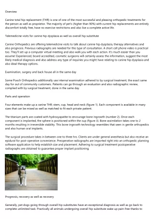
Folia Morphol., 2023, Vol. 82, No. 1 The musculocutaneous nerve (MCN) begins at the level of the inferior border of the pectoralis minor muscle. Following the classical manuals, it arises as a terminal branch from the lateral cord of the bra- chial plexus and passes through the coracobrachialis, then between biceps brachii and brachialis muscles to supply them. After that, it continues as “the lat- eral cutaneous nerve of the forearm”, which is the cutaneous innervation along the lateral side of the forearm. The branch to brachialis muscle supplies also the elbow joint. So that the MCN is responsible for motor innervations of the muscles of the anterior compartment of the arm and sensory supply to the skin of the lateral side of the forearm [20, 31]. Isolated MCN injuries have been diagnosed and reported in a variety of clinical situations, including direct trauma to the anterior shoulder, fractures of the humerus and clavicle, abundant fracture callous formation, anterior shoulder dislocations, gunshot wounds, lacerations, and intravenous catheterisation, also, some cases of MCN palsy were reported after forceful exercise [21, 34]. The nerve is at risk both with open and arthroscopic procedures (especially anterior shoulder surgery) and can be stretched by retractor placement on the coracobrachialis muscle for exposure [13]. So, attention should be taken in shoulder surgeries (e.g. shoulder joint replacement), before placing a retractor on the medial side of the incision to retract the conjoined muscles and pecto- ralis major, it is essential to identify the MCN to avoid it injury [22]. Variations of the MCN and its branches are com- mon; these variations have been described in human by many authors [14, 16, 23, 27, 29]. This study was conducted to demonstrate the anatomical variations in the origin, course, distribution, and branching pat- tern of the MCN in the axilla and arm and to define the intercommunications with the median nerve (MN) in the human male adult cadavers to prevent lesions during surgical procedures. Figure 1. The left axilla and arm showing the normal origin of mus- culocutaneous nerve (MCN): it arises from the lateral cord of the brachial plexus and piercing the coracobrachialis muscle (cb); MN — median nerve; UN — ulnar nerve; MC — medial cord. obtained from the dissection room of the Anatomy Department, Faculty of Medicine. The brachial plexus was dissected carefully with special concern to the exposure and topographic lo- calisation of the MCN regarding the variations of its origin, course, and branching pattern. These findings were photographed using a digital camera (Canon- -EOS-650D, made in Japan). In addition, a Vernier calliper was used to measure the length of the MCN and its branches. The MCN was traced from the cora- coid process to the lateral epicondyle of the humer- us. The MCN was studied as regards its branches, distribution, and communication with other nerves especially the MN. The branches arising from the MCN to innervate the biceps and brachialis muscles were identified and studied regarding their number, site of exit, length, and variations. Various univariate analyses were used to assess each variation, to clarify some of the relationships between the variables. All data were analysed using SPSS version 23. RESULTS Forty upper limbs of 20 cadavers were studied, the brachial plexus was dissected, and the MCN was studied on both right and left upper extremities. Several variations in the course and the branching pattern of the MCN were observed. MATERIALS AND METHODS This study was conducted after the approval from the Unit of Biomedical Ethics Research Committee in Faculty of Medicine, King Abdulaziz University, Saudi Arabia. All methods and techniques used dur- ing carrying out the research were in accordance with the protocol approved above. The present study was carried out on 40 upper limbs of 20 male adult cadavers fixed in 10% formalin. Preserved cadavers Origin of musculocutaneous nerve In 38 (18 right, 20 left) (95%) out of 40 upper limb specimens, the MCN was appearing from the lateral cord of the brachial plexus as described in the classical manuals (Fig. 1), in only one arm (right) 80
M.G. Al-Sobhi et al., Musculocutaneous nerve for surgical issues: anatomical study Figure 2. The right axilla and arm showing, the musculocutaneous nerve (MCN) arising from the lateral root (LR) of the median nerve (MN). MCN gives 3 branches; 1 to brachialis (br), 2 to short head of biceps (S), and 3 to long head (L) and then continues as lateral cutaneous nerve of the forearm (LCN). Additional branch to L from nerve to brachialis muscle (arrow). Figure 3. The right axilla and arm showing, the absence of mus- culocutaneous nerve. The median nerve (MN) gives 2 branches; 1 supplying biceps (B) muscle and 2 supplying brachialis (bbr) muscle and then continues as lateral cutaneous nerve of the fore- arm (LCN). Lateral cord (LC) gives (bcb) branch to coracobrachialis muscle. Table 1. Location of branches of musculocutaneous nerve in the arm Right arm 29.17 ± 2.45 7.71 ± 1.23 26.39 ± 3.22 12.14 ± 2.56 41.29 ± 6.24 Left arm 29.31 ± 2.15 7.78 ± 2.01 26.47 ± 5.88 12.72 ± 2.18 43.44 ± 6.93 Coracoid-lateral epicondyle distance [cm] Average distance from coracoid process to coracobrachialis muscle [cm] Average distance from coracoid process to coracobrachialis muscle as % of coracoid-lateral epicondyle distance Average distance from coracoid process to emergence of nerves supplying biceps brachii muscle [cm] Average distance from coracoid process to emergence of nerve(s) supplying biceps brachii muscle as % of coracoid-lateral epicondyle distance Average distance from coracoid process to emergence of nerves supplying brachialis muscle [cm] Average distance from coracoid process to emergence of nerve(s) supplying brachialis muscle as % of coracoid-lateral epicondyle distance Average distance from coracoid process to emergence of communicating branch to median nerve [cm] Average distance from coracoid process to emergence of communicating branch to median nerve as % of coracoid-lateral epicondyle distance 15.38 ± 3.39 52.34 ± 8.62 17.19 ± 3.93* 58.15 ± 11.04* 11.73 ± 4.13 42.22 ± 8.43 13.07 ± 2.79 44.47 ± 8.36 Data are shown as mean ± standard deviation. Student t-test: *p < 0.05 compared to the right side. (2.5%) MCN arose from the MN (Fig. 2), while in the remaining arm (right) (2.5%) it was absent (Fig. 3). Regarding the coracoid-lateral epicondyle distance it was approximated in both right (29.17 ± 2.45 cm) and left (29.31 ± 2.15 cm) upper limbs (Table 1). part of the muscle (Fig. 8). While in the remaining four specimens (3 right, 1 left) (10%) the MCN did not enter the coracobrachialis muscle, in this case an isolated branch originated from the lateral cord of the brachial plexus and pierced the coracobrachialis muscle to supply it instead of the MCN (Figs. 2, 9). Average distance (cm) from coracoid process to cora- cobrachialis muscle was approximated in both right (7.71 ± 1.23) and left (7.78 ± 2.01) arms, repre- senting a percentage of coracoid-lateral epicondyle distance equal to 26.39 ± 3.22 in right arm and 26.47 ± 5.88 in left arm (Table 1). Biceps and brachialis muscles. In 35 (15 right, 20 left) (87.5%) out of 40 specimens, the branches of the MCN that innervate the biceps and brachialis muscles arose from it after it leaves the coracobra- chialis muscle (Fig. 4). In 5 specimens (right) (12.5%) Relations of musculocutaneous nerve with the muscles of the arm Coracobrachialis muscle. In 26 (14 right, 12 left) (65%) of the specimens, the MCN entered the upper part of coracobrachialis muscle (Fig. 4), while in four (2 right, 2 left) (10%) it entered its middle part (Figs. 5, 6), in another four (1 right, 3 left) (10%) it entered the lower part of the muscle (Fig. 7), and in two specimens (left) (5%) it entered the upper part of the coracobrachialis muscle and gives a branch to biceps muscle, then the main trunk entered again the lower 81
Folia Morphol., 2023, Vol. 82, No. 1 Figure 4. The right axilla and arm showing, the musculocutaneous nerve (MCN) entered the superior part of the coracobrachialis mus- cle (cb). It gives branch 1 which bifurcates to supply biceps (B) muscle. It gives also communicating branch (arrowhead) with the median nerve (MN). Figure 7. The left axilla and arm showing, the musculocutaneous nerve (MCN) gives 3 branches (a) to biceps (bs), to brachialis (b) and communicating (C) with the median nerve (MN); UN — ulnar nerve; Cb — coracobrachialis. Figure 8. The left axilla and arm showing, the musculocutaneous nerve (MCN) gives branch 1 to supply biceps muscle (bs) muscle and communicating branch (C) to join the median nerve (MN) which gives (B1) to supply brachialis (br) muscle. The main trunk of MCN gives (B2) to supply also br muscle, then continues as lateral cuta- neous nerve of the forearm (LCN); cb — coracobrachialis muscle. Figure 5. The left axilla and arm showing, the musculocutaneous nerve (MCN) gives a communicating branch (C) which pierces coracobrachialis (cb) muscle to join the median nerve (MN). MCN supplies brachialis muscle (br) by only one branch (arrow). Figure 9. The right axilla and arm showing, the musculocutaneous nerve (MCN) joins the median nerve (MN) by a short trunk (arrow). MCN gives branch 1 to biceps (B) andbranch to (bbr) brachialis (br) muscle and continues as lateral cutaneous nerve of the forearm (LCN); UN — ulnar nerve. Figure 6. The right axilla and arm showing, the musculocutane- ous nerve (MCN) gives (bb) branch which bifurcates into a and b branches to supply short (S) and long (L) heads of biceps muscle and gives also 1, 2, 3 to supply brachialis muscle (br) then contin- ues as lateral cutaneous nerve of the forearm (LCN); cb — coraco- brachialis muscle. 82
M.G. Al-Sobhi et al., Musculocutaneous nerve for surgical issues: anatomical study Table 2. Length of musculocutaneous nerve branches in the arm muscle is shorter; statistically non-significance; in the right (12.14 ± 2.56) than the left (12.72 ± 2.18) arms, repre- senting a percentage of coracoid-lateral epicondyle dis- tance which is also shorter; statistically non-significance; the right (41.29 ± 6.24) than that in the left (43.44 ± ± 6.93) arms (Table 1). Regarding the length of branches to the short head of biceps brachii it is shorter; statistically non-significance; in the left (3.22 ± 1.02) than in the right (3.47 ± 0.91) arms (Table 2). Also, the length of branches to the long head of biceps brachii is shorter; statistically non-significance; in the left (3.88 ± 1.36) than in the right (4.27 ± 1.23) arms (Table 2). So, the length of branches to short head of biceps brachii are shorter than that of long head without statistically significance difference (Table 2). Two anatomical variations were observed for the innervation of the biceps brachii muscle in this study: — type 1: a solar branch from the musculocutaneous that is divided to supply the two heads of the biceps muscle individually; seen in 33 (14 right, 19 left) (94.3%) of studied arms; — type 2: in two limbs (1 right, 1 left) (5.7%), two separate branches arose from the musculocutane- ous, one to supply the long head while the other one to supply the short head of the biceps. There was an additional branch innervating the distal part of the long head of biceps (Fig. 2). Patterns of the branches supplying the brachialis muscle (35 specimens). The average distance (cm) from the coracoid process to emergence of motor branches innervating the brachialis muscle is shorter; with statistically significant difference p < 0.05; in the right (15.38 ± 3.39) than the left (17.19 ± 3.93) arms, representing a percentage of coracoid-lateral epicon- dyle distance which is also shorter; with statistically sig- nificant difference p < 0.05; in the right (52.34 ± 8.62) than that in the left (58.15 ± 11.04) arms (Table 1). Regarding the length of branches to brachialis muscle it is shorter; statistically non-significance; in the left (5.04 ± 1.2) than in the right (5.35 ± 1.86) arms (Table 2). Three types of anatomical variations were observed: — type I: it is found in 29 specimens (19 right, 10 left) (82.9%) of arms, where there was a single branch innervating the brachialis muscle from the main trunk of MCN (Fig. 9); — type II: in 5 specimens (4 right, 1 left) (14.2%) of arms, there were two branches that innervate the brachialis muscle from the main trunk of MCN (Figs. 10, 11); Length of motor branch supplying Long head of biceps brachii muscle Short head of biceps brachii muscle Brachialis muscle Right arm 4.27 ± 1.23 3.47 ± 0.91 5.35 ± 1.86 Left arm 3.88 ± 1.36 3.22 ± 1.02 5.04 ± 1.21 Data are shown as mean ± standard deviation. Figure 10. The right axilla and arm showing, the musculocutaneous nerve (MCN) gives branch 1 which bifurcates to supply short (S) and long (L) heads of biceps muscle and branches 2 and 3 to supply brachialis (br) muscle; cb — coracobrachialis muscle; LCN — lateral cutaneous nerve of forearm. Figure 11. The right axilla and arm showing, the musculocutane- ous nerve (MCN) divided into two branches: 1 — branch to biceps and 2 — branch continue as lateral cutaneous nerve of forearm (LCN) and give a and b branches to brachialis (br) and communi- cating branch (C); MN — median nerve. the branches to both biceps and brachialis muscles, along with the lateral cutaneous nerve of the forearm, arose from the MN itself (Figs. 2, 3). Distribution of musculocutaneous nerve Patterns of the branches supplying the biceps brachii muscle (35 specimens). The average distance (cm) from the coracoid process to emergence of motor branches to both short and long heads of biceps brachii 83
Folia Morphol., 2023, Vol. 82, No. 1 (in 8 specimens) and joined the distal part of the MN (in other 4 specimens) (Figs. 5, 7, 11). DISCUSSION Embryologically, the limb buds are developed from the lateral plate of the mesoderm and the mes- enchyme of those buds discriminate into the deep structures of the limbs, whereas the axons of the peripheral nerves develop in a distal direction from the ectoderm to reach the muscles and skin [11]. The somite migration led to formation of the extremities, where they bring their own nerve supply, so every dermatome and myotome keeps the original seg- mental innervation. During somite migration, some of the nerves come into close proximity and fuse in a particular pattern, forming a plexus early in fetal life [1, 2]. The existence of anatomical neuromus- cular variations maybe due to different factors that enhance the pathway of muscle formation in the limbs. Factors guiding nerve growth are chemo-at- tractive and repellent that control cellular prolif- eration to proper tissue formation. Butz et al. [6] stated that signalling mechanisms during embry- ogenesis could have a role during the 5th week of gestation, the axons of spinal nerves propagate distally to reach the mesenchyme of the limb, and insufficient signalling may negatively impact the normal formation of the brachial plexus. This em- bryological clarification justifies what we observed in our finding. The anatomical variations from the expected pat- tern of peripheral nerve course and relations can be a challenge for the surgeons. In the arm, variations of the nerves that innervate the anterior compartment (musculocutaneous, median, and ulnar nerves) are more common than those of the posterior compart- ment [7, 25]. Musculocutaneous nerve is a terminal branch of the brachial plexus, which provides the chief motor innervation for the arm flexors besides the sensory innervation for the lateral side of the forearm. In the present study, MCN originated from the lateral cord of the brachial plexus in 90% of cases while from the MN in only 5% of cases. These findings were in agreement with Bergman et al. [4], who reported that this nerve arose from the lateral cord in 90.5%, but on the contrary to this study they found that the MCN arose from the MN in only 2% of specimens. Moreover, they reported that it might be doubled, unusually short or absent. Figure 12. The right axilla and arm showing, the musculocutane- ous nerve (MCN) gives a communicating branch (C) to join the me- dian nerve (MN) before piercing the coracobrachialis muscle (cb); UN — ulnar nerve. — type III: in one right specimen (2.9%) of arms, three branches innervating the brachialis muscle, these branches originated also from the main trunk of MCN (Fig. 6). Patterns of communication between muscu- locutaneous and median nerves. This communica- tion was observed in 24 (11 right, 13 left) (60%) out of 40 specimens. The average distance (cm) from the coracoid pro- cess to emergence of communicating branch to MN is shorter; statistically non-significance; in the right (11.73 ± 4.13) than the left (13.07 ± 2.79) arms, representing a percentage of coracoid lateral epi- condyle distance which is also shorter; statistically non-significance; the right (42.22 ± 8.43) than that in the left (44.47 ± 8.36) arms (Table 1). The communicating branches were categorised based on their origin from the MCN and its union with the MN: There are three different types of communications: — type A: the proximal part of the MCN sharing a common trunk with the proximal part of the MN. This finding was observed in 4 specimens (3 right, 1 left) (16.7%) (Fig. 9); — type B: the proximal part of the MCN gives a commu- nicating branch to join the middle part of the MN, it was observed in 6 specimens (2 right, 4 left) (25%) (Fig. 12). In two arms (1 right, 1 left) (8.3%), of the previous specimens, a branch arising from this com- munication supplying the brachialis muscle (Fig. 8); — type C: in the remaining 12 specimens (4 right, 8 left) (50%), within the coracobrachialis muscle, a communicating branch arose from the middle part of the MCN to join the middle part of the MN 84
M.G. Al-Sobhi et al., Musculocutaneous nerve for surgical issues: anatomical study In the present study, the MCN was found to be absent in 5% of cases. The absence of the MCN was reported by many authors in previous studies [27, 29]. In particular, a case study was similar to the pres- ent study, in that the motor nerve to the coraco- brachialis muscle arose from the lateral cord, while the motor nerve to the biceps brachii and brachialis muscles arose from the MN [15]. Variable pathways and relations of the MCN with- in the coracobrachialis muscle were described. Ozturk et al. [24] stated that the MCN pierced the coracobra- chialis muscle in all studied 42 specimens of upper limb, whereas Pacha Vicente et al. [26] and Eglseder and Goldman [10] observed that the MCN did not enter the coracobrachialis muscle in 29.6% and 6.5% of their samples, respectively. Furthermore, Macchi et al. [19] observed a range of differences in the entry site of the MCN into the coracobrachialis muscle, and that was correlated with a low variability in the exit site of the nerve from the muscle. However, the exit point was positively related to the length of the muscle. Choi et al. [8] stated that the MCN penetrated the coracobrachialis muscle at a lower level in a single arm but did not pierce it in 4.7% of the specimens. Uysal et al. [32] observed that the MCN pierced the upper part of the coracobrachialis muscle in 43% of studied limbs and its middle part in 37% and its lower part in 17%, while it did not pierce it in only 3% of samples. In the present study, the MCN pierced the upper part of coracobrachialis muscle in 65% of the specimens, while in only 10%, it pierced its middle part and in another 10% it pierced its lower part. In only 5% of specimens, it entered the upper part of the coracobrachialis muscle and gives a branch to biceps muscle then the main trunk entered again the lower part of the muscle. While in the remaining 10% of specimens, the MCN did not penetrate the muscle. These observations demonstrated the relations between the MCN and the coracobrachialis muscle. Furthermore, it shows the probability of nerve injury particularly when the upper and middle parts of the coracobrachialis muscle are exposed to trauma. Earlier studies revealed the appearance of the MCN using ultrasound. Schafhalter-Zoppoth and Gray [30] observed that if this nerve was not visible in the coracobrachialis muscle, it was probably fused with the MN, later it is separated from it. The MCN inner- vates the coracobrachialis and the biceps brachii, plus the majority of brachialis muscle. The branch supply- ing the coracobrachialis arose from the MCN prior to piercing the muscle, while the branches supplying the biceps and brachialis muscles originating from it after its exit from the muscle [18, 30]. The significance of the nerves supplying the bi- ceps and brachialis in the surgeries of the brachial plexus has been extensively acknowledged [26]. In a previous study on the branches of the MCN to both biceps and brachialis muscles; it was stated that there are three types of the innervation pattern to biceps muscle observed in 24 studied cadavers [35]. Type I, one main branch arose from the main trunk of the MCN distal to the coracobrachialis muscle and con- sequently divided into two branches to supply each (short and long) heads of the biceps muscle. Type II, two main branches for each head of the biceps separately, the proximal branch for the short head and the distal one for the long head of the muscle. Type III, two main branches; a proximal branch gives two subdivisions, each one to supply a head of the biceps muscle, plus a distal one to supply the com- mon belly. A study found type I in 83.3% of cases while in this study it was found in 95.5% of cases [35], while in Pacha Vicente’s findings, it was 60.5% [26]. Types II and III were stated in 8.3% and 8.3% of cases respectively by Yang et al. [35], while it was 27.9% and 11.6% respectively by Pacha Vicente et al. [26]. In 5% of specimens in this study, there was no example found of type III branching pattern to the biceps that was defined by Elgammal et al. [12], which was three isolated main branches: first to long head, second to short head and the third one to the common belly. The measurements that were carried on the exit point of the first branch to biceps and brachialis muscles are specified in past reports [12]. In this study the methodology described by Yang et al. [35] and Elgammal et al. [12] was applied, which uses the coracoid process of the scapula and the medial epicondyle of the humerus as reference points for these measurements was followed. Yang et al. [35] observed two innervaion patterns of the brachialis muscle (type I showing one main branch, and type II showing two main branches). The type II innervation pattern was demonstrated in 8.4%, in the present study, but it was 4.2% of the samples of Yang et al. [35] while 27.9% of the samples of Pacha Vicente [16, 26, 35]. An additional type to those described by Yang et al. [35] was seen in a single specimen (5%) in this study, where there were three branches innervating the brachialis muscle, 85
Folia Morphol., 2023, Vol. 82, No. 1 these branches originated also from the main trunk of MCN [9, 14, 35]. Intercommunications between the MCN and MN had an important clinical significance, especially in relative to the accurate explanation of clinical neu- rophysiology, realizing the anatomy of the anterior shoulder repairs after trauma, and recognizing the dysfunction of median and MCNs [8, 9]. The fre- quency of these communications has been reported to differ between 5% and 46.4% [23]. Interestingly, a case was reported in a cadaver showing that the MCN gives a third root to form the MN [6]. Although intercommunicating branches most commonly origi- nate from the MCN and joined the MN, both report- ed incidents where the intercommunicating branch originates from the MN and joined the MCN [18, 26]. In the present study, the reverse was observed; the intercommunicating branch arose from the MCN and joined the MN in 35% of arms. Similar to Uysal et al. [32], a branch originating from the communicating branch between the MCN and the MN to the brachialis was seen in this study in only a single arm [16, 32]. Choi et al. [8] detected that in 26.4% of cases, there was communicating branches or fusion of the MCN and MN [8, 16]. The communicating branches were classified into three patterns; 1st pattern (19.2%) revealed merging of the MCN and MN, 2nd pattern (74%) had one branch communicating between the MCN and MN, while 3rd pattern (6.8%) had two branches communicating between the two nerves. It has been also described that the communicating branch originated from the MCN proximal to the entry point to the coracobrachialis muscle in 29.4%, through the muscle in 2%, distal to it in 54.9%, and from it as the MCN did not enter the muscle in 13.7% of cases [8, 14]. On the other hand, Venieratos and Anagnost- opoulou [33] studied 22 specimens and they catego- rised the communicating branches into three types: proximal to the entry point of the musculocutaneous nerve into the coracobrachialis in 9 specimens (type I), distal to coracobrachialis in 10 specimens (type II), and beyond coracobrachialis in 3 specimens (type III) [23, 33]. In this work, the communicating branches between the MCN and MN were observed and cat- egorised into three types according to where they arose and merged the respective nerves. Type A — the proximal part of the MCN sharing a common trunk with the proximal part of the MN in only one specimen (14.2%), type B — the com- municating branch arose from the proximal part of the MCN to merge the middle part of the MN in two (28.4%) specimen and in type C (57.2%), the most observed type, the communicating branch arose from the middle part of the MCN within the coracobra- chialis muscle to merge the middle part of the MN, these observation was in agreement with findings of Ballesteros et al. [3]. These finding were in the contrary to Nascimento et al. [21] who stated that the point of joining the MCN with the MN is distal to the coracobrachialis muscle in type II, and type III, where neither the nerve nor the communicating branch pierce the coracobrachialis muscle [3, 5]. The site of nerve communication and the number of branches that originate from the MCN to merge the MN may change the clinical symptoms and case progression along with management. Therefore, these differences should be considered during the clinical examination and treatment of traumatic injuries to upper limb. CONCLUSIONS The presented data in our work that demonstrate the branches of the MCN are significant for surgical doctors who perform operational procedures in the axilla and upper arm region. Acknowledgements The authors wish to express their gratitude to all those who donated their bodies to medical science so that anatomical research could be performed. Results from such research can potentially increase mankind’s overall knowledge that can then improve patient care. Therefore, these donors and their families deserve our highest gratitude [17]. Conflict of interest: None declared REFERENCES 1. Afshar A. An update on embryology of the upper limb. J Hand Surg Am. 2013; 38(11): 2304, doi: 10.1016/j. jhsa.2013.09.018, indexed in Pubmed: 24207000. 2. Al-Qattan M, Kozin S. Update on embryology of the upper limb. J Hand Surg. 2013; 38(9): 1835–1844, doi: 10.1016/j. jhsa.2013.03.018. 3. Ballesteros LE, Forero PL, Buitrago ER. Communication between the musculocutaneous and median nerves in the arm: an anatomical study and clinical implications. Rev Bras Ortop. 2015; 50(5): 567–572, doi: 10.1016/j. rboe.2014.08.009, indexed in Pubmed: 26535190. 4. Bergman RA, Afifi AK, Miyauchi R. Illustrated Encyclo- pedia of Human Anatomic Variation. Opus III: Nervous System. 2015. 86
M.G. Al-Sobhi et al., Musculocutaneous nerve for surgical issues: anatomical study 5. Budhiraja V, Rastogi R, Kumar Asthana A, et al. Concurrent variations of median and musculocutaneous nerves and their clinical correlation--a cadaveric study. Ital J Anat Em- bryol. 2011; 116(2): 67–72, indexed in Pubmed: 22303635. 6. Butz JJ, Shiwlochan DG, Brown KC, et al. Bilateral variations of brachial plexus involving the median nerve and lateral cord: An anatomical case study with clinical implications. Australas Med J. 2014; 7(5): 227–231, doi: 10.4066/ AMJ.2014.2070, indexed in Pubmed: 24944720. 7. Chelly JE. Peripheral nerve blocks: a color atlas. Lippincott Williams & Wilkins 2009. 8. Choi D, Rodríguez-Niedenführ M, Vázquez T, et al. Patterns of connections between the musculocutaneous and median nerves in the axilla and arm. Clin Anat. 2002; 15(1): 11–17, doi: 10.1002/ca.1085, indexed in Pubmed: 11835538. 9. Chrysikos D, Athanasopoulos A, Georgakopoulos P, et al. Anatomical variation of a communicating branch between the musculocutaneous and the median nerve: a case re- port. Acta Med Acad. 2020; 49(1): 71–74, doi: 10.5644/ ama2006-124.286, indexed in Pubmed: 32738120. 10. Eglseder WA, Goldman M. Anatomic variations of the mus- culocutaneous nerve in the arm. Am J Orthop (Belle Mead NJ). 1997; 26(11): 777–780, indexed in Pubmed: 9402212. 11. El-Naggar MM, Al-Saggaf S. Variant of the coracobrachialis muscle with a tunnel for the median nerve and brachial artery. Clin Anat. 2004; 17(2): 139–143, doi: 10.1002/ ca.10213, indexed in Pubmed: 14974102. 12. Elgammal Y, Frolov A, Martin J. The branching pattern and internal topography of the musculocutaneous nerve. FASEB J. 2020; 34(S1): 1–1, doi: 10.1096/fasebj.2020.34.s1.04481. 13. Frazer EA, Hobson M, McDonald SW. The distribution of the radial and musculocutaneous nerves in the brachialis muscle. Clin Anat. 2007; 20(7): 785–789, doi: 10.1002/ ca.20521, indexed in Pubmed: 17854055. 14. Gelmi C, Pedrini F, Fermi M, et al. Communication between median and musculocutaneous nerve at the level of cubital fossa: a case report. Transl Res Anat. 2018; 11: 1–4, doi: 10.1016/j.tria.2018.04.001. 15. Gümüsburun E, Adigüzel E. A variation of the brachial plexus characterized by the absence of the musculocu- taneous nerve: a case report. Surg Radiol Anat. 2000; 22(1): 63–65, doi: 10.1007/s00276-000-0063-x, indexed in Pubmed: 10863751. 16. Hunter D, Zdilla M. The absent musculocutaneous nerve: A systematic review. Transl Res Anat. 2021; 22: 100092, doi: 10.1016/j.tria.2020.100092. 17. Iwanaga J, Singh V, Takeda S, et al. Acknowledging the use of human cadaveric tissues in research papers: Recommenda- tions from anatomical journal editors. Clin Anat. 2021; 34(1): 2–4, doi: 10.1002/ca.23671, indexed in Pubmed: 32808702. 18. Krishnamurthy A, Nayak SR, Venkatraya Prabhu L, et al. The branching pattern and communications of the musculocuta- neous nerve. J Hand Surg Eur Vol. 2007; 32(5): 560–562, doi: 10.1016/J.JHSE.2007.06.003, indexed in Pubmed: 17950223. 19. Macchi V, Tiengo C, Porzionato A, et al. Musculocutaneous nerve: histotopographic study and clinical implications. Clin Anat. 2007; 20(4): 400–406, doi: 10.1002/ca.20402, indexed in Pubmed: 17022027. 20. Moore KL, Dalley AF. Clinically oriented anatomy. Wolters Kluwer india Pvt Ltd 2018. 21. Nascimento SR, Ruiz CR, Pereira E, et al. Rare anatomical variation of the musculocutaneous nerve: case report. Rev Bras Ortop. 2016; 51(3): 366–369, doi: 10.1016/j. rboe.2015.08.019, indexed in Pubmed: 27274492. 22. Occhiboi E, Clement R. Anatomic total shoulder arthroplasty and reverse total shoulder arthroplasty. JBJS J Orthop Physi- cian Assist. 2020; 8(1): 0025, doi: 10.2106/jbjs.jopa.19.00025. 23. Orellana-Donoso M, Valenzuela-Fuenzalida J, Gold- Semmler M, et al. Neural entrapments associated with musculoskeletal anatomical variations of the upper limb: Literature review. Transl Res Anat. 2021; 22: 100094, doi: 10.1016/j.tria.2020.100094. 24. Ozturk A, Bayraktar B, Taskara N, et al. Morphometric study of the nerves entering into the coracobrachialis muscle. Surg Radiol Anat. 2005; 27(4): 308–311, doi: 10.1007/ s00276-005-0326-7, indexed in Pubmed: 15968480. 25. Oztürk NC, Uzmansel D, Oztürk H. An unreported pattern of musculocutaneous and median nerve communication with multiple variations of biceps brachii: a case report. Surg Radiol Anat. 2010; 32(9): 887–890, doi: 10.1007/ s00276-009-0616-6, indexed in Pubmed: 20049596. 26. Pacha Vicente D, Forcada Calvet P, Carrera Burgaya A, et al. Innervation of biceps brachii and brachialis: Anatomical and surgical approach. Clin Anat. 2005; 18(3): 186–194, doi: 10.1002/ca.20057, indexed in Pubmed: 15768419. 27. Pacholczak R, Klimek-Piotrowska W, Walocha JA. Absence of the musculocutaneous nerve associated with a supernu- merary head of biceps brachii: a case report. Surg Radiol Anat. 2011; 33(6): 551–554, doi: 10.1007/s00276-010- 0771-9, indexed in Pubmed: 21225427. 28. Pandey SK, Shukla VK. Anatomical variations of the cords of brachial plexus and the median nerve. Clin Anat. 2007; 20(2): 150–156, doi: 10.1002/ca.20365, indexed in Pubmed: 16795062. 29. Parchand MP, Patil ST. Absence of musculocutaneous nerve with variations in course and distribution of the median nerve. Anat Sci Int. 2013; 88(1): 58–60, doi: 10.1007/ s12565-011-0126-6, indexed in Pubmed: 22237923. 30. Schafhalter-Zoppoth I, Gray AT. The musculocutaneous nerve: ultrasound appearance for peripheral nerve block. Reg Anesth Pain Med. 2005; 30(4): 385–390, doi: 10.1016/j. rapm.2004.12.008, indexed in Pubmed: 16032591. 31. Snell RS. Snell’s Clinical Anatomy. Wolters Kluwer India Pvt Ltd. 2018. 32. Uysal II, Seker M, Karabulut AK, et al. Brachial plexus variations in human fetuses. Neurosurgery. 2003; 53(3): 676–684, doi: 10.1227/01.neu.0000079485.24016.70, indexed in Pubmed: 12943583. 33. Venieratos D, Anagnostopoulou S. Classification of com- munications between the musculocutaneous and median nerves. Clin Anat. 1998; 11(5): 327–331, doi: 10.1002/ (SICI)1098-2353(1998)11:5<327::AID-CA6>3.0.CO;2-M, indexed in Pubmed: 9725577. 34. Vineyard AP, Gallucci AR, Imbus SR, et al. Residents case re- port: musculocutaneous nerve injury in a collegiate baseball pitcher. Int J Sports Phys Ther. 2020; 15(5): 804–813, doi: 10.26603/ijspt20200804, indexed in Pubmed: 33110700. 35. Yang ZX, Pho R, Kour AK, et al. The musculocutaneous nerve and its branches to the biceps and brachialis mus- cles. J Hand Surg Am. 1995; 20(4): 671–675, doi: 10.1016/ s0363-5023(05)80289-8. 87