Download

1 / 11
0 likes | 1 Views
Background<br>Short morning exposure to high illuminance visible electromagnetic radiations termed as artificial daylight is beneficial for the mental health of people living in geographical areas with important seasonal changes in daylight illuminance. However, the commercial success of high illuminance light sources has raised the question of the safety of long hour exposure.<br>Methods<br>We have investigated the effect of the replacement of natural daylight by artificial daylight in Swiss mice raised under natural lighting conditions. Mice were monitored for neurotoxicity and general health changes

E N D
Journal of Microscopy and Ultrastructure 5(2017) 206–215 Contents lists available at ScienceDirect Journal of Microscopy and Ultrastructure jou rnal hom epage: www.elsevier.com/locate/jmau Original Evaluation artificial Article of the safety of conventional lighting replacement by daylight Eteta,∗, Farahnaa,b, Khayrc,d, Omare, Paul Ömür Süleyman F. Seke Mohammed Maher A.M. Khalid M. Denizf, Mustafag,Nadia Alattad, Alhayanig, G. Hesham N. O. Abdulmonem Kaplanf, Vecchioa Lorella aDepartment bDepartment Khartoum, cDepartment dFaculty eDepartment fDepartment gDepartment ofBasic Health Sciences, College of Applied Medical Sciences, Qassim University, 51452 Buraydah, Saudi Arabia of Anatomy, Faculty of Medicine and Health Sciences, Al-Neelain University, Development and Innovation Center, Tabil Food Industries, Sudan of Physics, Faculty of Education, Alzaeim Alazhari University, Khartoum North, Sudan of Radiological Sciences and Nuclear Medicine, National Ribat University, Khartoum, Sudan of Physics, Faculty of Science and Arts, Almethnab, Qassim University, Saudi Arabia ofHistology and Embryology, Medical Faculty, Ondokuz Mayis University, 55139, Samsun, Turkey ofAnatomy, Faculty of Medicine, King Abdulaziz University, Jeddah, Saudi Arabia a r t i c l e i n f o a b s t r a c t Article Received Accepted Available history: Short morning exposure to high illuminance visible electromagnetic radiations termed as Background: artificial seasonal sources Methods: Swiss health functions’ signs entorhinal Results: observed to entorhinal the Conclusions: radiations damages hour ©2017 10 April 2017 daylight is beneficial for the mental health of people living in geographical areas with important 14 May 2017 changes in daylight illuminance. However, the commercial success of high illuminance light online 1June 2017 has raised the question ofthe safety of long hour exposure. We have investigated the effect of the replacement of natural daylight by artificial daylight in Keywords: Entorhinal Hippocampus Bright Behavior Mood Mouse mice raised under natural lighting conditions. Mice were monitored for neurotoxicity and general cortex changes. They were submitted to a battery of conventional tests for mood, motor and cognitive assessment on exposure day (ED) 14 and ED20.Following sacrifice on ED21 due to marked visible light of neurotoxicity, the expression of markers of inflammation and apoptosis was assessed in the cortex and neurons were estimated in the hippocampal formation. disorder Signs of severe cognitive and motor impairments, mood disorders, and hepatotoxicity were in animals exposed to artificial daylight on ED20, unlike on ED14 and unlike groups exposed natural daylight or conventional lighting. Activated microglia and astrocytes were observed in the cortex, as well as dead and dying neurons. Neuronal counts revealed massive neuronal loss in hippocampal formation. These results suggest that long hour exposure to high illuminance visible electromagnetic induced severe alterations in brain function and general health in mice partly mediated by to the neocortex-entorhinal cortex-hippocampus axis. These findings raise caution over long use ofhigh illuminance artificial light. Saudi Society of Microscopes. Published by Elsevier Ltd. This isan open access article under the CC BY-NC-ND license (http://creativecommons.org/licenses/by-nc-nd/4.0/). 1. Introduction graphical latitudes, ocular cal accompanied disorder [1–5].Typically, sive energy centration, Laboratory and seasonal changes, particularly Arctic and Antarctic and winter. These changes result inalterations of the Light exposure is apowerful environmental cue for the reg- diurnal rhythms, sleep-wake cycle, and other biologi- ulation daytime of circadian rhythms inmammals. Important changes in rhythms, including the neuroendocrine and immune system, duration and daylight illuminance associate with geo- by serious health problems like seasonal affective (SAD), with ahigher frequency of occurrence inwomen SAD patients display symptoms of major depres- disorders, including psychomotor retardation, agitation, and ∗Corresponding Applied Arabia. E-mail author at: Department of Basic Health Sciences, College of loss, anhedonia, indecisiveness, decreased interest and con- Medical Sciences, Qassim University, P.O. Box 6699, 51452 Buraydah, Saudi feelings of worthlessness, and suicidal ideation [6–9]. rodents are also affected byseasonal changes, justify- paul.seke@gmail.com (P.F. Seke Etet). address: http://dx.doi.org/10.1016/j.jmau.2017.05.005 2213-879X/© licenses/by-nc-nd/4.0/). 2017 Saudi Society of Microscopes. Published by Elsevier Ltd. This isan open access article under the CC BY-NC-ND license (http://creativecommons.org/
207 P.F. Seke Etet et al. /Journal of Microscopy and Ultrastructure 5(2017) 206–215 ing [10–12]. Artificial exposure treatment sonal daylight being, and thermore, improved disease abilities eliciting Together of totherapy artificial ing radiations tial present light functions, their use inmechanistic studies of SAD and related conditions detect determined On battery cognition, a.m. inrooms dure was mined tests, The increases ply under lected was processed tion Embryology, the labeling sis Abdulaziz important changes insocial behavior. The body weight was every three days. light positive properties at specific illuminance and exposure days (ED) 14 and 20, mice were submitted toa time allowed the development of phototherapy for the of behavioral tests aimed atassessing changes inthe mood, of mood and biological disorders associated with sea- and motor function. Tests were performed between 11 changes. Daily exposure (morning, upto2-h) toartificial (Zeitgeber 5, 5hafter light phase onset) and 2p.m. (ZT 8), i.e. (bright white artificial light >8000 lx) improved well- where animals were housed. The whole testing proce- sleep, daytime psychomotor vigilance performance, cortisol required 30min per animal. The performance of each animal melatonin levels, and SAD patients’ condition [13–18].Fur- video recorded and scored offline. Skin temperature was deter- strategic exposure toartificial daylight during daytime inboth ears at the beginning and the end ofthe battery of SAD-like primary and secondary features of Parkinson’s using anon-contact infrared thermometer. [19],but also learning effectiveness and other cognitive experiment inlive animals was stopped onED21 due to inpeople affected by seasonal changes [20,3,21],without inthe aforementioned signs of toxicity, inorder tocom- any major safety concerns [22,23]. with animal research ethical standards. Animals were sacrificed with other benefits of bright light and the low cost deep gas anesthesia between ZT5and ZT8. Blood was col- the technology, the aforementioned positive effects of pho- bycardiac puncture and brains dissected out and fixed. Blood contribute tothe commercial success of daylight-grade processed for liver function test. Brains’ left hemispheres were light sources. Surprisingly, considering notably emerg- for histopathological studies and stereological estima- evidence supporting adverse effects of related electromagnetic of hippocampal neurons at the Department of Histology and such as UVA [24,25],noreport is available on the poten- Ondokuz Mayis University (Samsun, Turkey). Instead, adverse reactions tolong exposure toartificial daylight. In the right hemispheres were processed for immunohistochemical study, we assessed the impact of continuous artificial day- of resident cells and markers of inflammation and apopto- exposure during daytime on the physiology, mood, cognitive inthe entorhinal cortex, at the Department of Anatomy, King and number of cells inthe brain of mice. University (Jeddah, Saudi Arabia). 2.1.3. Artificial Artificial system homogeneous Department ing 3.3 tance daylight exposure daylight was delivered by alinear source lamp (LSL) 2. Methods designed and optimized toproduce isothermal, regular, and electromagnetic radiation by Qassim University’s 2.1. Animals and procedures of Physics [26].The system was made of a focus- mirror and eight cool white fluorescent tubes (60 cmlength, 2.1.1. Animals Swiss from College bia). divided (70 room cage conventional (21.736 on iments Arabia, time) the at (9152B, ature food The approved of performed of and light exposure cmdiameter, 1800 lm) inthe same horizontal plane. The dis- female mice (n =24) were raised under natural daylight, between the LSL system and the cage floor was 60cm. birth tothe age of8months, inthe animal facility of the of Pharmacy, Qassim University (Buraydah, Saudi Ara- 2.2. Behavioral tests Then, these animals (36.87 3.83 gweight) were randomly ± in three groups (n =8) housed intransparent Plexiglas cages The following tests were performed sequentially: cm×70cm, height 60 cm) indifferent rooms. Acage was ina exposed tonatural daylight (22.000 lxaverage luminance at 2.2.1. Footprint The ment. an walls) consecutive exclude test floor), while the other two were inrooms either exposed to footprint test was performed for gait and balance assess- lighting (500 lxatcage floor) ortoartificial daylight Mice with inked paws were allowed towalk freely along lxatcage floor) during the daytime. Lights were switched enclosed box (70 cm long, 7cmwide, and 20 cmhigh plexiglas and off byan automated system, based on dawn and dusk. Exper- with a clean sheet ofpaper placed onthe floor. After three inlive animals were performed inBuraydah, Central Saudi tests, only one valid trial was considered per animal to during the winter, thus civilian dawn was at 6:24 a.m. (local habituation phase-associated abnormal patterns [27]. and dusk at6:11 p.m. (daytime length: ∼11h47 min). During dark phase (night), rooms were kept under dim red light (3lx 2.2.2. Elevated The two and was parameters eledlaced and occurred the episodes plus maze cage floor). Light intensity was monitored using aphotometer EPM consisted of two open arms (30 cm×7cm, nowall), Pasco Scientific, Roseville, CA). Ineach room the temper- closed arms (30 cm×7cm, with 20 cmhigh Plexiglas walls), was constant (23.5◦C) and animals had access to ad libitum acommon central platform (7cm×7cm). The entire apparatus and water. elevated to70 cmabove floor level. Each mouse was plogical experiment and all procedures inlive animals were scored included entries, time spent, and distance trav- byboth the Research Center and the Ethical Committee on the central platform of the maze, facing an open arm, the College of Applied Medical Sciences, Qassim University, and the behavior was recorded for 5min. Etho ineach arm. An entry according toECDirective 2010/63/EU on the protection when all four limbs were within an arm. Head dips over animals inscientific experiments. edge of open arms, rearing, grooming, sniffing, and freezing were also counted. 2.1.2. Experimental Animals artificial and vocalization (‘red (mainly reflexes). erized procedures exposed tonatural daylight, conventional lighting, or Open Exploratory Plexiglas divided (remaining started activity era 2.2.3. field test daylight were monitored daily, todetect signs of systemic behavior was examined inan open field arena in central nervous system toxicity such as shaggy fur, cachexia, (40.6 cm×40.6 cm, height 38.1 cm). The arena’s floor was when handled, and porphyrin deposits around the eye into acentral (20.2 cm×20.2 cm) and a peripheral zone tears’). Negative geotaxis and various reflexes were asserted 10.2 cmsurrounding the central zone). The test was posture, pinna, righting, contact righting, and corneal byplacing amouse inthe arena, facing the wall. Animal’s Animal behavior incages was recorded with acomput- inthe chamber was recorded for 10 min using acam- system equipped with infrared cameras and analyzed to mounted on side (approximately 50 cm from the floor) that
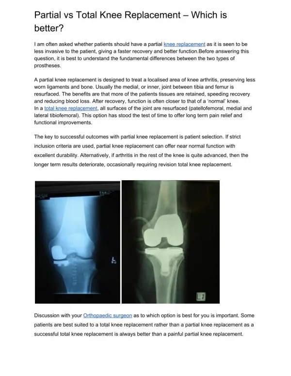
208 P.F. Seke Etet et al. /Journal of Microscopy and Ultrastructure 5(2017) 206–215 captured tor computing tracking eled as both vertical and horizontal activities. Mouse locomo- technique program (coefficient oferror ≤0.05) applied using the relevant activity was tracked with an application developed inMATLAB ofthe stereological workstation. environment (MathWorks, Natick, MA) using motion 2.4.2. Immunohistochemistry Standard inimage sequences. Entries, time spent, and distance trav- procedures were used, as previously described [31], inthe central and peripheral zones were determined, as well on embedded antibodies 1:1000 goat TNF-? (apoptosis receptor, apoptosis, lated Santa chromogen was were observed pus (Olympus, randomly selected sections (6?m-thick) made on paraffin- the time spent inthe corners. brain right hemispheres, inthe coronal plane. Primary used were goat anti-NeuN (mature neuron marker, Y-maze The mended 30 between and placed LAB used total per another An 2.2.4. spontaneous alternation test dilution), goat anti-GFAP (astrocyte marker, 1:100 dilution), test was performed under reduced light (∼100 lx), as recom- anti-iba1/CD68 (microglial marker, 1:1000 dilution), goat anti- [28,29].Y-maze consisted of three Plexiglas arms (length (pro-inflammatory cytokine, 1:500 dilution), rabbit anti-Fas cm, width 7cm, height 20 cm) symmetrically placed (120◦angle mediator, 1:500 dilution), rabbit anti-Fas ligand (death arms). Each mouse was placed inthe center of the maze 1:500 dilution), and rabbit anti-caspase 3(marker of spontaneous activity was recorded for 5min using a camera 1:500 dilution) (Santa Cruz Biotechnology, CA). Biotiny- above the maze. The following were determined using MAT- anti-goat IgG and biotinylated anti-rabbit IgG (1:200 dilution, applications: the number and sequence of arm entries, the time Cruz Biotechnology, CA) were the secondary antibodies. The toperform the first three and the last three alternations, the substrate 3,3?-diaminobenzidine hydrochloride (DAB) distance traveled, and the average speed (distance traveled applied and counterstained with hematoxylin. Glass coverslips time unit). Direction changes occurring after sniffing the wall of placed using DPX mounting medium. Sections labeled were arm (hesitation during arm change) were also determined. with acomputerized light microscope including an Olym- entry occurred when all four limbs were within the arm. BX53 microscope equipped with an Olympus DP73 camera Tokyo, Japan), under 4×,20×, 40× and 100×objectives. 2.3. Liver function test 2.5. Data analysis Levels of total protein, albumin, globulin, triglycerides and cholesterol, aminotransferase determined Laboratory facturer’s of lated. as well as the activities of the enzymes aspartate One way ANOVA for independent groups followed by LSD test (AST) and alanine aminotransferase (ALT) were was group ioral test between ferences presented used toassess the statistical significance of inter-treatment insera using commercially available kits (bio-Merieux changes inbody weight, body temperature, and behav- Reagents and Products, France), according tothe manu- test parameters. Repeated measures ANOVA followed byLSD instructions. The albumin/globulins ratio and the fraction was used toassess the statistical significance ofdifferences total protein represented byalbumin and globulins were calcu- ED14 and ED20 performances inbehavioral tests. Dif- with p-value lower than 0.05 were significant. Data are as mean ±SEM. 2.4. Tissue processing and histopathological studies 3. Results Brains were sequentially fixed in10% formalin for 8-h, abun- dantly soaked don rinsed inPBS, dried inincreasing concentrations of ethanol, 3.1. Animal general condition inxylene, and embedded inparaffin using Thermon Shan- Citadel tissue processor (GMI Inc., Ramsey, MN, USA). Neurological From more expressed in mals all These exploratory visual more tion. However, group; was observed not 3.1.1. examination ED8 onward, artificial daylight–exposed animals were Histopathology Sections throughout with a dom Selected drated, then, phology Stereological ator equipped BrieldField; areas mined bright 4×). The ing most dentate tively, the objective gyrus 2.4.1. and stereology agitated when handled than animals of other groups and of embedded tissues (20 ?m-thick) were made audible vocalizations. Besides, fights were more frequent the entire left brain hemisphere inthe coronal plane, the cage of artificial daylight–exposed animals. Thus, three ani- arotary microtome (Leica RM2125RT) using steel blades. After presented with bite injuries on the back onED10, and almost random selection of the first section, the rule of systematic ran- presented with such injuries on legs, feet, and the back onED20. sampling with 1/5 ratio was applied tothe section selection. animals also displayed shaggy and dirty furs, decreases in sections were mounted on slides, deparaffinized, rehy- activity, regular freezing episodes, as well as slower stained with cresyl violet (Nissl staining), dehydrated, and placing and negative geotaxis responses. They also resisted mounted with Entellan using standard procedures. The mor- tothe separation from grid during the grip strength evalua- of neurons in the hippocampal formation was examined. analyses were performed using optical fraction- no major locomotor impairment was observed inany method [30] and acomputerized stereological workstation grip strength (measured by the ability tohold tight toagrid) with StereoInvestigator software (version 9.0, Micro- also comparable between groups; and no marked change was Colchester; USA). Briefly, before starting the counting, inthe reflexes assessed, inthe lacrimation (the eyes were (coordinates) of CA1, CA2, CA3, and dentate gyrus were deter- dry), and inthe salivation (no dry mouth). on every section and for each animal using an atlas and a field microscope with motorized stage (microscope objective 3.1.2. Body The weight in, ventional y=1.17x increase 0and group polynomial weight effect and artificial body temperature daylight These areas of interest were scanned along the x-and y-axes. of exposure on the body x/y grid sizes, the step size, and the area of unbiased count- isshown inFig. 1A. Body weight increased linearly frame atmicroscope objective 100×were determined (i.e. the and was comparable between, natural daylight- and con- R2=0.97 appropriate step and frame toavoid counting CA1–CA3 and lighting–exposed groups (y=1.02x +36.1, and R2=0.98, gyrus neurons twice). Afterwards, pyramidal cells (respec- +35.6, respectively). However, acomparable granular cells) were counted inCA1–CA3 areas (respectively, was observed inartificial daylight group only between ED R2=0.99). dentate gyrus) using the unbiased counting frame (microscope ED4(y=1.75x +34.88, Afterwards, animals of this 100×).Then, total numbers of neurons inthe dentate displayed atransient decrease inbody weight following a (y=0.58x2−3.53x R2=0.94), and CA1–CA3 were estimated with the optical fractionator progression +41.43, with
209 P.F. Seke Etet et al. /Journal of Microscopy and Ultrastructure 5(2017) 206–215 Fig. A. in daylight exposed walking behavioral traveled the with mean 1.Body weight and behavioral tests. Body weight progression. Note the non-linear and lower increase inartificial daylight-exposed animals. B. Body temperature before and after test battery. Note the decrease temperature atthe end of tests inartificial daylight group on both exposure day (ED) 14and 20. C–F. Footprints of representative cases of animals exposed tonatural (C) and conventional lighting (D) onED20, and animals exposed toartificial daylight on ED14 (E) and 20 (F). Note the regular pattern of steps innatural daylight (D), the slightly irregular steps inconventional lighting-exposed (E), and the decreased ground contact surface of hind paws (oval shapes) and incoordination of compared with both natural daylight and conventional lighting groups (arrows) inartificial daylight-exposed (F). G–L. ED 14 (light gray) and EG 20(dark grey) tests’ results. G, H. Distance traveled inthe central zone of the open field arena (G) and central zone latency (H). Note the significant decrease inthe distance inthe central zone and increase inthe latency inartificial light-exposed group. I,J.Time spent inelevated plus maze open arms (I) and closed arm latency (J). Note significant decreases compared tonatural daylight-exposed group. K, L. Average speed atthe first six entries inY-maze arms (K) and number of arm changes made hesitation (L). Note the decrease inspeed and increase inarm changes with hesitation inartificial daylight-exposed group more marked onED20 than ED 14. Data are ±SEM. ANOVA +LSD test: *P<0.05, ** P<0.01, *** P<0.001. arestorative weight lower, (Fig. Theeffect test’s ature conventional pared the group inflection point on ED10 (Fig. 1A). The average body tery values p=0.033 ing, test (p=0.03 on ED14, p=0.01 on ED20) and with post-battery of the group exposed toartificial daylight was significantly of other groups (ED14: p=0.01 conventional lighting, vs. compared tothe other groups, from ED10 onward (p=0.034) vs. natural daylight/ED20: p=0.02 vs. conventional light- 1A). p=0.028 natural daylight) (Fig. 1B). vs. of artificial daylight exposure on pre- and post-battery body (skin) temperature is shown inFig. 1B. Body temper- Serum The proteins, increases Total cial 3.1.3. molecules reflecting hepatic function before the battery ofbehavioral tests was slightly higher in effect of artificial daylight exposure on levels of serum lighting- and artificial daylight–exposed groups com- lipids, and hepatic enzymes isshown inTable 1.Slight tonatural daylight–exposed. Body temperature at the end of intotal cholesterol and triglyceride levels were observed. behavioral tests was significantly lower inartificial daylight protein, albumin, and globulin levels were higher inartifi- on both ED14 and ED20 compared with values before bat- daylight–exposed group, compared with natural daylight- and,
210 P.F. Seke Etet et al. /Journal of Microscopy and Ultrastructure 5(2017) 206–215 Table1 Effect of light source/intensity onlevels of serum proteins and molecules reflecting hepatic function. rangesa natural daylight conv. lighting artificial daylight ANOVA: F; pvalues (Fcrit =4.6) Reported nat. Dvs. Conv. L nat. Dvs. art. D Conv. Lvs. art. D Serum Total Albumin % Globulin % A/G protein levels (g/L) 70.5 39.4 55.9 29.4 41.5 1.4 protein ±1.2 74 41.3 55.8 24.5 33 1.8 ±0.5↑ 79.6 47.8 60 38.8 48.8 1.2 ±0.5↑ 8;0.025* 0.9; 0.001; 2.4; 4.5; 0.14; 52; 12.4; 1.65; 22; 10; 0.31; 0.0001*** 68;0.0001*** 20; 15.2; 20; 15; 0.047; 67 46.5 38 29.5 38 1.7 ±9 1.9 ±0.8 1.4↑ 0.36 0.008** 0.002** ±7.8 ± ± of total ±2.8 ±2.8 ±1.7↑ 1 0.235 0.005** ±9.8 ±1.3 ±1.3 ±1.5↑ 0.16 0.0016** 0.002** ±6.4 of total ±6 ±3.7 ±1.7↑ 0.06 0.012* 0.005** ±9.8 ratio ±0.26 ±0.53 ±0.06 2.711 1.13 5.49* ±0.34 Hepaticenzymes ALT AST T. Triglycerides and lipid 68.7 29 125 231 levels (mg/dL) (IU/L) ±39.2 71.5 39.6 121 225 ±36.4 186.6 67.4 118 248 ±63.1 0.91; 0.16; 0.1; 0.01; 0.013 12.6; 8.11; 0.001; 0.06; 0.008** 12.5;0.007** 0.05; 0.04; 0.5; 60 38 110 150 ±13.2 (IU/L) ±13.5 ±7.6 ±27 2.315 0.021* 5.96 ±21.9 cholesterol ±60 ±60 ±80 0.81 0.97 0.85 ±29 ±63 ±63 ±31 0.9 0.81 0.51 ±42 aRanges ***P reported by recent studies performed under normal laboratory lighting inmice [33,20,37,3].T. cholesterol: total cholesterol. ANOVA +LSD test: *P<0.05; **P < 0.01; <0.001. Data are mean ±SEM. inalesser these (Table significantly ventional Serum (AST) cial conventional light literature extent, conventional lighting–exposed. The levels of and grooming light (Table relative time, rearing (on hind legs and against wall), and proteins were also higher than physiological values [32–34] latency were significantly decreased inartificial day- 1).Albumin/globulins ratio inartificial daylight group was group, while freezing and sniffing episodes were increased lower (respectively, slightly lower) compared tocon- 2). lighting (respectively, natural daylight). levels of the liver enzymes aspartate aminotransferase 3.2.3. Elevated The The inartificial lighting–exposed natural ED20) inartificial lighting– daylight–exposed Closed shorter ED20) Rearing conventional (p on cial conventional (p central freezing cial alesser and (grooming plus maze and alanine aminotransferase (ALT) were higher inartifi- results of EPM test are shown inFig. 1I,Jand Table 2. daylight–exposed animals compared tonatural daylight- and time spent inmaze open arms was significantly decreased lighting–exposed. Levels of AST and ALT inartificial daylight–exposed group compared with conventional group were higher than physiological values reported inthe (p=0.001 on ED14 and p=0.0012 on ED20) and [32–34] (Table 1). daylight–exposed (p=0.0051 on ED14 and p=0.0029 on (Fig. 1I). Closed arm latency was significantly decreased daylight–exposed group compared with conventional 3.2. Behavioral tests (p=0.052 on ED14 and p =0.037 on ED20) and natural (p=0.0051 on ED14 and p=0.0023 on ED20). Footprints’ Representative conventional Artificial marked and compared also tional to compared 3.2.1. analysis cases of animals exposed tonatural daylight, arm latency of conventional lighting–exposed was also lighting, and artificial daylight are shown inFig. 1C–F. compared with natural daylight–exposed (p=0.037 on daylight–exposed animals displayed static gait alterations (Fig. 1J). byadecreased size of contact (more obvious inhind paws) against wall frequency was significantly increased in anincoordination of walking (gait ataxia) on ED20 (Fig. 1F) lighting- (p=0.018) and inartificial daylight–exposed with their walking performance on ED14 (Fig. 1E), and =0.002) on ED14 compared with natural daylight. Instead, compared with both natural daylight- (Fig. 1C) and conven- ED20 adecrease inthis parameter was observed inartifi- lighting–exposed (Fig. 1B). Notably, some animals exposed daylight–exposed compared with ED14 (p=0.0001), but also conventional lighting presented slightly irregular step patterns lighting– (p=0.025) and natural daylight–exposed with animals exposed tonatural daylight. =0.007) (Table 2). As also shown inTable 2,open arm entries, platform time, and head dips over the open arms, and episode frequency were significantly decreased inartifi- Open Open distance in and (Table was daylight–exposed both and zone than the significantly compared groups lighting–exposed) icantly other vs p 3.2.2. field daylight–exposed group compared tonatural daylight, and in field test results are shown inFig. 1G, Hand Table 2.The extent, conventional lighting. On the other hand, fecal boli traveled inthe open field arena was significantly decreased episodes of rearing, undirected sniffing, and grooming attempts artificial daylight–exposed group on both ED14 (p=0.002) episodes shorter than 3s)were increased (Table 2). ED20 (p=0.0059) compared with natural daylight–exposed 2). The distance traveled inthe central zone of the arena significantly lower inconventional lighting- and artificial Y-maze Y-maze average daylight–exposed and ral with entries hesitation another group (p=0.0003 daylight–exposed) was ural 3.2.4. compared with natural daylight–exposed on test results are shown inFig. 1K, Land Table 2.The ED14 (p=0.014 and p=0.009, respectively) and ED20 (p=0.03 speed inthe first six entries was decreased inartificial p=0.009, respectively). The distance traveled inthe central group on ED20 compared with ED14 (p= 0.008) by artificial daylight–exposed was also significantly lower toconventional lighting–exposed (p=0.001), but not natu- conventional lighting–exposed (p=0.048) (Fig. 1G). Time to daylight–exposed (Fig. 1K). Asimilar scenario was observed first entry in the central zone (central zone latency) was the speed difference between the first six and the last six higher inartificial daylight–exposed group on ED20 (Table 2). The frequency of episodes of arm change with with ED14 (p=0.042), but compared also to the other (sniffing of an arm entrance followed by the choice of (p=0.014 natural daylight– and p =0.03 vsconventional arm) was significantly higher inartificial daylight–exposed vs. (Fig. 1H). The time spent incorners was signif- on ED20 compared with ED14 (p=0.003) and toother groups higher inartificial daylight–exposed compared with the conventional lighting–exposed, p =0.0001 natural vs. vs. groups on ED14 (p=0.0013 vs. natural daylight, p=0.0034 (Fig. 1L). Arm change with hesitation episodes conventional lighting) and ED20 (p=0.005 natural daylight, more common inconventional lighting–exposed than innat- vs. =0.039 vsconventional lighting) (Table 2). Central zone entries daylight–exposed (p=0.01) (Fig. 1L).
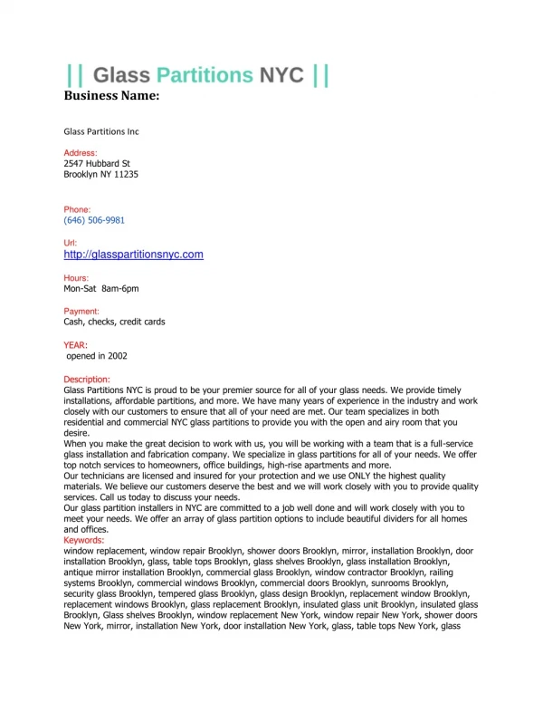
211 P.F. Seke Etet et al. /Journal of Microscopy and Ultrastructure 5(2017) 206–215 Table Effect 2 of light exposure on animal performance inbehavioral tests. Natural daylight Conventional lighting Artificial daylight ED14 ED20 ED14 ED20 ED14 ED20 Open Total Central Central Time Rearing Rearing Undirected Wall Floor Grooming, Grooming Freezing field distance, cm 2930 13.4 22 119.1 26.2 41.1 24±3 34.1 13.1 5.1 191.8 1.5 ±250 2561 12.7 21.8 111.6 25.6 39.8 21.4 34.6 12.2 5.8 193 1.4 ±200 2072 18 15.1 156.8 32.6 51 34.9 48.1 20.8 7.3 85.8 1.6 ±197b↓ 2159 18.5 16.1 174.3 35.7 55.3 38.4 52.7 23.1 7.3 92 1.3 ±257 1801 11.2 13.7 257.3 38.1 51.3 51.3 47.4 41.9 6.6 84.1 4.1 ±190b↓ 1607 5.9 11.9 246.9 18.4 40.7 62.6 77.3 67.3 5.4 99.9 4.1 217b↓ ± zone time, %total ±2.1 ±2.2 ±3 ±3.4 ±2.1c↓ ±1.4a,b,c↓ zone entries, nr ±1.8 ±2 ±2.6b↓ ±2.3b↓ ±1.7b↓ ±2.9b↓ spent incorners, s ±26 ±28 ±22 ±29 ±26a,b↑ ±31a,b↑ on hind legs, nr ±5.7 ±6.3 ±6.3 ±6.8 ±13.8 ±5.5c↓ against wall, nr ±3 ±2.5 ±5.5 ±4.9b↑ ±8.3 ±6c↓ sniffing, nr ±1.6 ±4.4b↑ ±5.3b↑ ±8.7b↑ ±3.3b,c↑ sniffing, nr ±3.1 ±2.9 ±4b↑ ±4b↑ 6.4b↑ ±5.9a,b,c↑ ± sniffing, nr ±1.3 ±1.1 ±2.3b↑ ±3b↑ ±3.1b,c↑ ±4.5a,b,c↑ nr ±1.4 ±1.4 ±1.5 ±1.6 ±1.5 ±1.2 latency, s ±44 ±38.5 ±23b↓ ±23.4b↓ ±4.3b↓ ±15b↓ episodes, nr ±1 ±0.7 ±0.5 ±0.5 ±1.5 ±0.9b,c↑ Elevated Distance, Total Open Central Head Rearing Rearing, Sniffing, Grooming Freezing, Fecal plus maze cm 411 13.1 51.6 21.9 18.1 5.7 3.5 18.2 5.6 2.7 1.2 ±60 420 13.4 50.6 22.2 18.6 6.2 4±1.8 18.2 5.8 2.8 0.8 ±65 544 15.3 51.8 32.3 37.3 13.5 14.4 40.9 4.6 2.1 1.3 ±61 527 15.1 50.9 25.9 34.3 14.4 14.1 44.6 5.8 1.8 1.9 ±58 469 13.1 17 31.1 15.3 17.4 14.4 63.4 3.7 3.6 1.9 ±45 390 7.9 19.5 18.4 13.9 7.1 11.3 62.3 6.6 4.3 2.3 55 ± arm entries, number ±2.8 ±3.1 ±2.7 ±1.7 ±1.9 ±2.0a,c↓ arm entries, %total ±8.9 ±9.9 ±5.4 ±5.6 ±2.7b,c↓ ±4.9b,c↓ platform time, s ±2.4 ±2.6 5.3 5.1 5.2c↓ ±4.3c↓ ± ± ± dips over open arm, nr ±2.5 ±2.7 ±5b↑ ±6.4b↑ ±1.9c↓ ±2.7c↓ against wall, nr ±2.0 ±2.0 ±2.1b↑ ±1.9b↑ ±2.0b↑ ±1.2a,c↓ nr ±1.7 ±4b↑ ±3.1b↑ ±3.4b↑ ±2.6a,c↓b↑ nr ±2.2 ±2.5 ±3.8b↑ ±4.6b↑ ±8.1b,c↑ ±5.7b,c↑ attempts, nr ±1.0 ±1.1 ±1.1 ±0.9 ±0.6 ±0.8a↑ nr ±1.1 ±1.1 ±0.5 ±0.7 ±1.0 ±0.6c↑ boli, nr ±0.3 ±0.3 ±0.5 ±0.6 ±0.9 ±0.7b↑ Y Distance, Alternation, X-Y-Z Speed maze cm 938 19.2 10.5 3.6 ±82 818 18.8 10 3.5 ±98 1330 26.6 15.4 6.1 ±168b↑ 1320 27.8 17.2 6.0 ±188b↑ 1349 24.3 15.0 6.6 ±137b↑ 1181 18.6 10.9 4.0 93b↑ ± nr ±1.4 ±1.6 ±2.7b↑ ±3.7b↑ ±3.1 ±1.8c↓ alternations, nr ±0.8 ±0.6 ±1.7b↑ ±2.4b↑ ±2.5 ±1.4c↓ first entries –last, cm/s ±1.1 ±1.2 ±0.4 ±0.7 ±0.9 0.5a,c↓ ± ED14, ED20: a,b,c: (a),vs. (b), (c). exposure days 14and 20. ANOVA +LSD test, respective exposure day 14 natural daylight and vs. conventional lighting Data are mean ±SEM. vs. The distance traveled inthe Y-maze was significantly shorter neuronal that of pared also tional with counts (percent of natural daylight counts) suggested in (p light) artificial of ventional (p cial conventional frequency ED20 (p natural daylight–exposed than inother groups on both ED14 the dentate gyrus was more affected than other structures = 0.012 conventional lighting, p =0.001 vs. artificial day- the hippocampal formation inartificial daylight–exposed com- vs. and ED20 (p=0.01 vs. conventional lighting, p=0.004 toconventional lighting–exposed (Fig. 2E). Relative counts vs. daylight) (Table 2).On the same hand, the frequency revealed adecrease (∼15%) inpyramidal neurons inconven- consecutive visits of three different arms was higher in con- lighting–exposed animals (but not ingranule cells) compared lighting–exposed group on ED14 (p=0.008) and ED20 natural daylight–exposed (Fig. 2E). =0.005) compared with natural –exposed, but on ED20 artifi- daylight–exposed displayed alower frequency compared with 3.3.2. cell Expressions of inflammation, apoptosis, and brain resident lighting–exposed (p=0.03). Similar, arm alternation markers The was also lower innatural daylight–exposed, except on expressions of markers labeled in the entorhinal cortex where adrop was observed inartificial daylight–exposed of conventional The (GFAP) pressed the cytes, neurons Also daylight–exposed the mediating marked and representative cases of animals exposed tonatural daylight, =0.02 conventional lighting). vs. lighting, and artificial daylight are shown inFig. 3. markers of activated microglia (iba-1) (Fig. 3A–C), astrocytes (Fig. 3D–F), and neurons (NeuN) (Fig. 3G–I) were overex- 3.3. Brain histopathological studies inmost artificial daylight–exposed animals, compared to other groups. Inaddition, the first displayed enlarged astro- 3.3.1. Neuronal The tion animals. Fig. cases ing, normal lighting–exposed in Results dentate dal in in ral counts morphology and counts microglia in activated macrophage shape, and dead (or dying) analysis of Nissl-stained neurons inthe hippocampal forma- (Fig. 3A–I). revealed marked neuronal loss inartificial daylight–exposed unlike the other groups, animals of the artificial This loss was particularly marked inthe dentate gyrus. group displayed increased expressions of: (i) 2A–C shows dentate gyrus granule cells of representative pro-inflammatory cytokine TNF-?(Fig. 3J–L); (ii) the receptor of animals exposed tonatural daylight, conventional light- cell-cell interaction-induced cell death associated with and artificial daylight. While dentate gyrus granule cells had inflammation FAS (Fig. 3M–O) and its ligand (Fig. 3P–R); shapes innatural daylight- (Fig. 2A) and conventional (iii) the marker of apoptosis caspase 3(Fig. 3S–U). (Fig. 2B) animals, these neurons were condensed artificial daylight–exposed (Fig. 2C). 4.Discussion of neuronal counts inCA1, CA2, and CA3, and inthe gyrus are shown inFig. 2D, E. Absolute counts of pyrami- neurons inCA1, CA2, and CA3 revealed significant decreases The results of the present study suggest systemic and central artificial daylight–exposed (P<0.001), and inalesser extent, nervous toartificial icant attempts system functional alterations inmice exposed exclusively conventional lighting–exposed animals compared with natu- daylight during daytime for 20days. Notably, signif- daylight–exposed (Fig. 2D). Similar results were obtained in decreases inthe frequency of rearing against wall, in i.e. of dentate gyrus granule cells (Fig. 2D). Relative values of toescape, inthe open field and the elevated plus maze
212 P.F. Seke Etet et al. /Journal of Microscopy and Ultrastructure 5(2017) 206–215 Fig. A–C. (C). (A, to group. 2. Hippocampal neuron observation and counts. Micrographs ofNissl-stained dentate gyrus neurons of representative cases of animals exposed tonatural daylight (A), conventional lighting (B), and artificial daylight Note the dead (black arrows) and the condensed cells (white arrows) inarficial daylight-exposed (C) and the normal shapes ofgranular cells inthe other groups (asterisks) B). D, E. Hippocampal neuron counts. Absolute (D) and relative (E) cell counts inthe CA1–CA3 (pyramidal cells) and dentate gyrus (DG, granular cells) of animals exposed natural daylight, conventional lighting, or artificial daylight during daytime for 20 days. Note the marked decreases inneuronal population inartificial daylight-exposed Data are mean ±SEM. (EPM), Y-maze unlike ing artificial tions depression the ing, exploratory The behavioral of increases ahallmark increased EPM were eled indicator close in number formed. increases dysfunction in cated decreases Y as well as drastic decreases inthe speed at first entries in decreased cial a We structure, cognitive Immunohistochemical cortex ence that the many death overexpressed, neurons observations in reological neurons get and observed, findings cortex-hippocampus neuroinflammation, alterations daylight Furthermore, health ficial ground contact surface of hind paws observed inartifi- arms were observed inartificial daylight group on ED20, daylight-exposed, compared with the other groups, indicated ED14 and unlike natural daylight and conventional light- motor dysfunction with central nervous system involvement. groups. These observations indicate that animals exposed to investigated the affection of amajor sensorimotor gating daylight developed coping deficit for the aversive situa- the entorhinal cortex, as apotential mechanism for the presented by the ethological tests [35,36].Other indicators of deficits induced by the exposure toartificial daylight. were observed inartificial daylight–exposed animals in labeling of resident cells inthe entorhinal third week, including: shaggy and dirty fur, decreased groom- of artificial daylight–exposed animals revealed the pres- increased aggressiveness, regular freezing episodes, decreased of enlarged astrocytes and brain macrophages, suggesting activity, and body weight loss. astrocytes and microglia were activated. On the same hand, performance of artificial daylight–exposed animals inthe pro-inflammatory cytokine TNF-?was overexpressed, and tests performed on ED20 also suggested other signs resident cells were positive for the inflammation-induced cognitive and motor impairment. Anxiety was suggested by receptor Fas and its ligand. Neuronal marker NeuN was insniffing episodes and by post-testing hypothermia, indicating that neurons were injured, and many ofmild chronic stress exposure [37,38].Inaddition, were positive for the marker of apoptosis caspase 3. These latency tofirst entry inthe anxiogenic areas of the suggested the presence of a marked inflammation (open arms) and of the open field arena (central zone) the entorhinal cortex associated with neuronal loss. Using ste- observed, together with decreases inthe distance trav- techniques, we estimated the number ofNissl-stained inthese areas. Moreover, thigmotaxis, astable and robust inthe hippocampal formation, amajor functional tar- of discomfort manifested by the tendency toremain of the entorhinal cortex. Losses indentate gyrus granule cells tothe walls (secure area) [39,40] was markedly increased as well as CA1–CA3 pyramidal neurons of hippocampus were artificial daylight–exposed, as indicated byincreases inthe and cell numbers were significantly decreased. These ofactivities close towalls inall the behavioral tests per- suggest that neuronal loss inthe neocortex-entorhinal Besides, ashift inbehavioral baseline was indicated by axis resulted atleast partly from detrimental inEPM central platform time [41].Prefrontal cortex and were among the drivers of brain functional was indicated bydecreased spontaneous alternations observed inthis study following replacement of natural the Y-maze [42,43].Spatial memory impairment was indi- with artificial daylight. byincreases inepisodes of arm change with hesitation and liver function test, performed toassess general in consecutive explorations of three different arms inthe status, suggested an affection of the hepatic function inarti- maze [42,44,45].Furthermore, the incoordination of walking and daylight–exposed animals. Marked increases inserum levels
213 P.F. Seke Etet et al. /Journal of Microscopy and Ultrastructure 5(2017) 206–215 Fig. Representative (D–F), artificial and 3.Entorhinal cortex immunolabeling. cases of natural daylight, conventional lighting, and artificial daylight-exposed animals. A–I. Brain resident cells: microglia (iba-1) (A–C), astrocytes (GFAP) and neurons (NeuN) (G–I). J–U. Markers of inflammation and apoptosis: TNF-?(J–L), Fas (M–O), Fas ligand (P–R), and caspase 3(S–U). Note the dying neurons inthe daylight-exposed (arrows inI,U), the increased expressions of TNF-?(L), Fas (O), Fas ligand (R), and caspase 3(U), as well as the marked activation of microglia (C) astrocytes (F). of albumin/globulins teins, well-established appears if brain aggravating observed Altogether, ficial brain. nitive light be mouse mild vation children the enzymes AST and ALT were observed, as well as decreased For ventional instance, teaching institutions should probably avoid using con- ratio, and abnormally high levels of total pro- lighting when natural daylight is available. albumin, and globulins. Considering that these alterations are indicators of high-grade hepatotoxicity [46–48],it 5.Conclusions that artificial daylight would have resulted inanimal death the exposure had continued. This finding suggests that liver-to- The present study addressed the consequences of replacing nat- signaling may also have accounted among the causative or ural general hepatotoxicity, ety, became the by and ronal cell observed ings daylight, daylight by either artificial daylight orconventional lighting for factors of the cognitive deficits and mood alterations health and cognition inmice. Mice presented with signs of inthe present study [49–51]. as well as increasing signs of neurotoxicity, anxi- our results suggest that long hour exposure toarti- depression, cognitive alterations, and motor impairment that daylight may have tremendous negative effects on mouse severe in the third week of exposure. The involvement of Interestingly, anearly study inhumans reported mild cog- neocortex-entorhinal cortex-hippocampus axis was suggested impairment following daytime exposure tobright artificial immunohistochemical studies that revealed neuroinflammation [52].Thus, artificial daylight exposure for long hours may neuronal loss inthe entorhinal cortex, but also by massive neu- detrimental for mental health inhumans as well. Alarmingly, loss in the hippocampal formation confirmed by stereological exposure toconventional lighting inour study induced a estimation. Mild cognitive and motor impairments were also cognitive deficit and motor impairment. Although this obser- inanimals exposed toconventional lighting. These find- should be verified inhumans, itraises caution over keeping suggest that the replacement of natural daylight byartificial indoor under conventional lighting during the daytime. and inalesser extent conventional lighting, had detri-
214 P.F. Seke Etet et al. /Journal of Microscopy and Ultrastructure 5(2017) 206–215 mental Our during exposure sensible effects on the brain function and general health inmice. Mott effects Springerplus [21] Outof conventional Int [22] Nurs [23] Southern [24] daylight artificial [25] UVA1phototherapy. [26] visible Exp. [27] quantitative Neurosci [28] et long-term deposition [29] CD73isamajor One2013;8:e66896. [30] total optical [31] kola diabetic 2017;195:159–65. [32] gigantea carcinoma [33] S, Med [34] Mota-Flores on EvidBased [35] Learned Brain [36] escape [37] Hypothermia postnatal [38] Theeffects emotional 2014;270:300–6. [39] behavior 2000;70:471–6. [40] open-field Psychol [41] antinociceptive upontreatment (Berl) [42] phencyclidine Basic [43] the TiO2 [44] anxiolytic danser [45] antagonists Rev [46] albumin nasopharyngeal [47] globulin 2014;177:671–8. [20] MS, Robinson DH, Williams-Black TH, McClelland SS. The supporting of high luminous conditions on grade 3oral reading fluency scores. findings raise concern over the extended use of artificial light 2014;3:53. the daytime, and call for studies assessing the intensity and Wahnschaffe A, Haedel S,Rodenbeck A, Stoll C, Rudolph H, Kozakov R, et al. time safe for humans, particularly for children, the most the lab and into the bathroom: evening short-term exposure to light suppresses melatonin and increases alertness perception. group inthe human population. JMol Sci 2013;14:2573–89. Howland RH. Somatic therapies for seasonal affective disorder. JPsychosoc Ment Health Serv 2009;47:17–20. Conflicts of interest statement Lane KL, Hovenic W, Ball K,Zachary CB. Daylight photodynamic therapy: the California experience. Lasers Surg Med 2015;47:168–72. Authors declare no conflict of interest. Lerche CM, Heerfordt IM, Heydenreich J, Wulf HC. Alternatives tooutdoor illumination for photodynamic therapy–use of greenhouses and light sources. Int JMol Sci 2016;17:309. Richer V, Lui H. Cross-sectional evaluation of acute adverse reactions during Acknowledgements Br JDermatol 2017, http://dx.doi.org/10.1111/bjd.15319. Abou-Zaid FA, Omar KHM, Salim EI, El-Deen AA, Bekhite MM. Can man made The present study was supported by the College of Applied Med- light radiation affect the reproductive capacity of male mice? Egypt J. ical Histology versity, University port Pharmacy, Sciences, Qassim University (Saudi Arabia), the Department of Biol 2006;2:81–91. Vandeputte C, Taymans JM, Casteels C, Coun F,Ni Y, Van LK, et al. Automated and Embryology, Medical Faculty of Ondokuz Mayis Uni- gait analysis inanimal models of movement disorders. BMC (Turkey), and the Department of Anatomy, King Abdulaziz 2010;11:92. (Saudi Arabia). Authors acknowledge the technical sup- Arendash GW, Gordon MN, Diamond DM, Austin LA, Hatcher JM, Jantzen P, al. Behavioral assessment of Alzheimer’s transgenic mice following of Prof. Osama H. Omer, and of the Colleges of Physics and Abeta vaccination: task specificity and correlations between Abeta Qassim University. and spatial memory. DNA Cell Biol 2001;20:737–44. Kulesskaya N, Voikar V, Peltola M, Yegutkin GG, Salmi M, Jalkanen S, et al. regulator of adenosinergic signalling inmouse brain. PLoS References West MJ, Slomianka L, Gundersen HJ. Unbiased stereological estimation of the number of neurons inthesubdivisions of the rat hippocampus using the Allebrandt et Chronobiol [2] 2012;29:379–94. [3] exposure secretion. [4] human [5] important [6] J,etal. ED: 2013;35:192–4. [7] ‘depression’ emotional (anhedonia) 2009;72:1–7. [8] upside-down. [9] et correlated peaks [10] underlying 2014;66:56–65. [11] rodent [12] melatonin BiolSci [13] Effects performance, 2013;30:988–97. [14] 2009;11:459–65. [15] bright psychomotor [16] cortical 2012;32:741–52. [17] Safety neonates. [18] studyof Acta [19] improve Chronobiol [1] KV, Teder-Laving M, Kantermann T, Peters A,Campbell H, Rudan I, fractionator. Anat Rec 1991;231(December (4)):482–97. al. Chronotype and sleep duration: the influence of season of assessment. Farahna M, Seke Etet PF, Osman SY, Yurt KK, Amir N, Vecchio L, etal. Garcinia Int 2014;31:731–40. aqueous suspension prevents cerebellar neurodegeneration inlong-term Arendt J.Biological rhythms during residence inpolar regions. Chronobiol Int rat –atype 1diabetes mellitus model. JEthnopharmacol Munch M,Linhart F,Borisuit A, Jaeggi SM, Scartezzini JL. Effects of prior light Habib MR, Karim MR. Effect of anhydrosophoradiol-3-acetate of Calotropis on early evening performance, subjective sleepiness, and hormonal (Linn.) flower asantitumoric agent against Ehrlich’s ascites Behav Neurosci 2012;126:196–203. inmice. Pharmacol Rep 2013;65:761–7. Roenneberg T, Kantermann T, Juda M, Vetter C, Allebrandt KV. Light and the Mostafavi SH, Fazilati M, Mostafavi SA, Vahhabi MR, Mostafavi F, Omidvarinia circadian clock. Handb Exp Pharmacol 2013:311–31. etal. Hepatotoxicity of Dorema aucheri (Bilhar) inalbino mice. Arch Iran Turner PL, Mainster MA. Circadian photoreception: ageing and the eye’s 2013;16:530–2. role insystemic health. Br JOphthalmol 2008;92:1439–44. Perez Gutierrez RM, Madrigales AD, Horcacitas MC, Garcia BE, Cruz VT, Belleville G, Foldes-Busque G, Dixon M, Marquis-Pelletier E, Barbeau S, Poitras JM. Ameliorative effect of hexane extract of phalaris canariensis Impact ofseasonal and lunar cycles onpsychological symptoms inthe high fat diet-induced obese and streptozotocin-induced diabetic mice. anempirical investigation of widely spread beliefs. Gen Hosp Psychiatry Complement Altern Med 2014;2014:145901. Chourbaji S, Zacher C, Sanchis-Segura C, Dormann C, Vollmayr B, Gass P. Charlton BG. Amodel for self-treatment of four sub-types of symptomatic helplessness: validity and reliability of depressive-like states inmice. using non-prescription agents: neuroticism (anxiety and Res Brain Res Protoc 2005;16:70–8. instability); malaise (fatigue and painful symptoms); demotivation Overmier JB, Seligman ME. Effects of inescapable shock upon subsequent and seasonal affective disorder ‘SAD’. Med Hypotheses and avoidance responding. JComp Physiol Psychol 1967;63:28–33. Mrdalj J,Lundegaard MA, Murison R, Konow JF, Milde AM, Pallesen S, et al. Lawrynowicz AE, Baker TD. Suicide and latitude inArgentina: Durkheim after chronic mild stress exposure inrats with a history of Am JPsychiatry 2005;162:1022. maternal separations. Chronobiol Int 2014;31:252–64. Postolache TT, Lapidus M, Sander ER, Langenberg P,Hamilton RG, Soriano JJ, Richter SH, Wollmann E,Schmidt M, Zillmann U, Hellweg R, Sprengel R, et al. al. Changes inallergy symptoms and depression scores are positively of neonatal cryoanaesthesia-induced hypothermia on adult inpatients with recurrent mood disorders exposed toseasonal behaviour and stress markers inC57BL/6 mice. Behav Brain Res inaeroallergens. Sci World J2007;7:1968–77. Ebling FJ. On the value of seasonal mammals for identifying mechanisms Blaszczyk JW, Tajchert K,Lapo I,Sadowski B. Acoustic startle and open-field the control of food intake and body weight. Horm Behav inmice bred for magnitude of swim analgesia. Physiol Behav Ferguson SA, Maier KL. Areview of seasonal/circannual effects of laboratory Leppanen PK, Ewalds-Kvist SB, Selander RK. Mice selectively bred for behavior. Physiol Behav 2013;119:130–6. thigmotaxis: life span and stability of the selection trait. JGen Rendon NM, Rudolph LM, Sengelaub DR, Demas GE. The agonistic adrenal: 2005;132:187–204. elicits female aggression via regulation of adrenal androgens. Proc Rodgers RJ, Lee C, Shepherd JK. Effects of diazepam on behavioural and 2015:282. responses tothe elevated plus-maze inmale mice depend Gabel V, Maire M, Reichert CF, Chellappa SL, Schmidt C, Hommes V, etal. regimen and prior maze experience. Psychopharmacology of artificial dawn and morning blue light ondaytime cognitive 1992;106:102–10. well-being, cortisol and melatonin levels. Chronobiol Int Gotesson J,Ericson M, Soderpalm B,Pickering C. Repeated ethanol but not impairs spontaneous alternation behaviour inthe Y-maze. Lewy AJ. Circadian misalignment inmood disturbances. Curr Psychiatry Rep Clin Pharmacol Toxicol 2012;110:347–52. Hu R,Gong X, Duan Y, Li N, Che Y, Cui Y, etal. Neurotoxicological effects and Phipps-Nelson J, Redman JR, Dijk DJ, Rajaratnam SM. Daytime exposure to impairment of spatial recognition memory inmice caused by exposure to light, as compared todim light, decreases sleepiness and improves nanoparticles. Biomaterials 2010;31:8043–50. vigilance performance. Sleep 2003;26:695–700. Harquin Simplice F,David ET, Herve Herve NA. Enhancing spatial memory: Rojas JC, Bruchey AK, Gonzalez-Lima F.Low-level light therapy improves and antidepressant effects of tapinanthus dodoneifolius (DC) metabolic capacity and memory retention. JAlzheimers Dis inmice. Neurol Res Int 2014;2014:974308. Rusu G, Popa G, Ochiuz L, Nechifor M, Tartau L. Effects of some dopamine Slusher TM, Vreman HJ, Olusanya BO, Wong RJ, Brearley AM, Vaucher YE, etal. on spatial memory performance inrats?experimental research. and efficacy of filtered sunlight intreatment of jaundice inAfrican Med Chir Soc Med Nat Iasi 2014;118:116–24. Pediatrics 2014;133:e1568–74. Du XJ, Tang LL, Mao YP, Sun Y, Zeng MS, Kang TB, etal. The pretreatment Yoshida A, Takagi A, Ikejima A, Takenaka H, Fukai T,Ikeda S. Aretrospective toglobulin ratio has predictive value for long-term mortality in 231 Japanese vitiligo patients with special reference tophototherapy. carcinoma. PLoS One 2014;9:e94473. Dermatovenerol Croat 2014;22:13–8. Jolles S,Borrell R, Zouwail S,Heaps A, Sharp H, Moody M, etal. Calculated Willis GL, Turner EJ. Primary and secondary features of Parkinson’s disease (CG) as ascreening test for antibody deficiency. Clin Exp Immunol with strategic exposure tobright light: acase series study. Int 2007;24:521–37.
215 P.F. Seke Etet et al. /Journal of Microscopy and Ultrastructure 5(2017) 206–215 [48] Mwanza Ultrasonographic bile [49] implications 2014;35:9–20. T, Miyamoto T, Okumura M, Kadosawa T, Fujinaga T. Datar ClinNeurol [51] failure. [52] morning Physiol [50] S, Wijdicks EF. Neurologic manifestations of acute liver failure. Handb evaluation of portal vein hemodynamics inexperimentally 2014;120:645–59. duct ligated dogs. Jpn JVet Res 1998;45:199–206. Shawcross DL, Wendon JA. The neurological manifestations of acute liver D’Mello C, Swain MG. Liver-brain interactions ininflammatory liver diseases: Neurochem Int 2012;60:662–71. for fatigue and mood disorders. Brain Behav Immun Lafrance C, Dumont M, Lesperance P, Lambert C. Daytime vigilance after bright light exposure involunteers subjected tosleep restriction. Behav 1998;63:803–10.