Download
1 / 46
460 likes | 482 Views
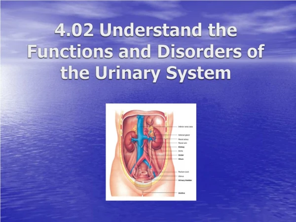
Explore the vital roles of the urinary system, from urine formation to blood composition regulation, and how it helps eliminate nitrogenous wastes and toxins. Learn about the structures of the kidneys and nephrons for effective urine filtration and reabsorption.

E N D
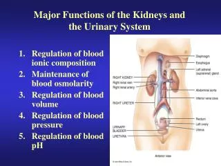
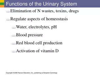
Functions of the Urinary System • Rinal diseas =09i8d • Nitrogenous wastes • Toxins • Drugs
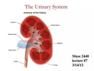
Blood Flow in the Kidneys Figure 15.2c
The structural and functional units of the kidneys • Responsible for forming urine • Main structures of the nephrons
A specialized capillary bed • Attached to arterioles on both sides (maintains high pressure) • Large afferent arteriole • Narrow efferent arteriole Figure 15.3c
Capillaries are covered with podocytes from the renal tubule • The glomerulus sits within a glomerular (Bowman’s) capsule (the first part of the renal tubule) Glomerulus Figure 15.3c
Renal Tubule Figure 15.3b
Located entirely in the cortex • Includes most nephrons Types of Nephrons Figure 15.3a
Found at the boundary of the cortex and medulla (loop of Henle dips into medulla) Types of Nephrons Figure 15.3a
Afferent arteriole feeds glomerular capillary bed • Efferent arteriole drains glomerular capillary bed • Arterioles are high resistance vessels • Afferent diameter is greater than Efferent diameter, so blood pressure in glomerulus is extremely high • The high BP forces fluids and solutes out of the blood and into the glomerular capsule that drains into the collecting tubule Glomerular Capillaries
Arise from efferent arteriole of the glomerulus • Normal, low pressure capillaries • Attached to a venule • Cling close to the renal tubule • Reabsorb (reclaim) some substances from collecting tubes Peritubular Capillaries
Urine Formation Processes Figure 15.4
Water and solutes smaller than proteins are forced through capillary walls • Blood cells cannot pass out to the capillaries • Filtrate is collected in the glomerular capsule and leaves via the renal tubule Filtration
Some water • Glucose • Amino acids • Ions • Some reabsorption is passive, most is active • Most reabsorption occurs in the proximal convoluted tubule (PCT) Reabsorption
Materials Not Reabsorbed • Nitrogenous waste products • Urea • Uric acid • Creatinine • Excess water
Secretion – Reabsorption in Reverse • Some materials move from the peritubular capillaries into the renal tubules • Hydrogen and potassium ions • Creatinine • Materials left in the renal tubule move toward the ureter
Formation of Urine Figure 15.5
Colored somewhat yellow (straw) due to the pigment urochrome (from the destruction of hemoglobin) and solutes • Sterile • Slightly aromatic • Normal pH of around 6 • Specific gravity of 1.001 to 1.035 Characteristics of Urine Used for Medical Diagnosis
= excessive sugar intake, diabetes mellitus • = physical activity, pregnancy, glomerulonephritis, hypertension • = urinary tract infection • = bleeding (kidney stone, infection) • = transfusion reaction, hemolytic anemia • = hepatitis Characteristics of Urine Used for Medical Diagnosis (Dip Stick)
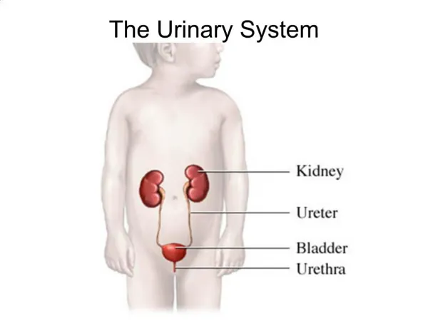
Ureters • Continuous with the renal pelvis • Enter the posterior aspect of the bladder • Runs behind the peritoneum • Peristalsis aids gravity in urine transport
Smooth, collapsible, muscular sac • Temporarily stores urine • Retroperitoneal, behind pubic symphysis Urinary Bladder Figure 15.6
– three openings • Two from the ureters • One to the urethrea Urinary Bladder Figure 15.6
Three layers of smooth muscle ( ) • Mucosa made of transitional epithelium • Walls are thick and folded in an empty bladder • In males the prostate gland surrounds the neck of the bladder where it empties into urethra Urinary Bladder Wall
Thin-walled tube that carries urine from the bladder to the outside of the body by peristalsis • Release of urine is controlled by two sphincters Urethra
Length • Females – 3–4 cm (1.5 inches) • Males – 20 cm (8 inches) • Location • Females – along wall of the vagina, opening anterior to vaginal opening • Males – through the prostate and penis Urethra Gender Differences
Function • Females – • Males – • 3 parts of urethra in males • – through prostate • – through membraneous tissue between prostatic and penile • – through penis Urethra Gender Differences
Both sphincter muscles must open to allow voiding • The internal urethral sphincter is relaxed after stretching of the bladder • Activation is from an impulse sent to the spinal cord and then back to bladder via the pelvic splanchnic nerves causing contractions • This sends urine past internal sphincter, and causes the sensation of having to void • The external urethral sphincter must be voluntarily relaxed
Blood composition depends on: • Diet, cellular metabolism, & urine output • In 24 hours the kidneys filter 150 – 180 liters of blood plasma, but only 1 – 1.8 liters of urine is produced • Filtrate is same as blood plasma, and as it moves along CD the kidney reabsorbs the good stuff (water, nutrients, ions), and secretes more of the bad stuff (nitrogenous wastes, and unneeded substances) • The left over filtrate is Urine Fluid, Elecrolyte, & Acid-Base Balance
Kidney’s role in blood composition: • 1) • 2) • 3) • 4) Fluid, Elecrolyte, & Acid-Base Balance
Normal amount of water in the human body • Young adult females – 50% (F>fat, F<musles) • Young adult males – 60% • Babies – 75% (low fat, low bone mass) • Old age – 45% • Water is necessary for many body functions and levels must be maintained Maintaining Water Balance
Interstitial fluid • Blood plasma • CSF, lymph, serous, eye humors, etc. Distribution of Body Fluid (Fluid Compartments) Figure 15.7
Changes in electrolyte balance (solute concentrations in/between compartments) causes water to move from one compartment to another • Alters blood volume and blood pressure • Can impair the activity of cells The Link Between Water and Salt
Sources for water intake • Ingested foods and fluids • Water produced from metabolic processes • Cellular metabolism (small amount) • Sources for water output • Vaporization out of the lungs • Lost in perspiration • Leaves the body in the feces • Urine production (will vary w/ kidney control) Maintaining Water Balance
Maintaining Water Balance • Dilute urine is produced if water intake is excessive • Less urine (concentrated) is produced if large amounts of water are lost • Likewise, proper concentrations of various electrolytes must be present
Regulation is primarily by hormones • prevents excessive water loss in urine • regulates sodium ion content of extracellular fluid • Triggered by the rennin-angiotensin mechanism • Cells in the kidneys and hypothalamus are active monitors Regulation of Water and Electrolyte Reabsorption
Maintaining Water and Electrolyte Balance Figure 15.9
Maintaining Acid-Base Balance in Blood • Blood pH must remain between 7.35 and 7.45 to maintain homeostasis • Most (hydrogen) ions originate as byproducts of cellular metabolism
Maintaining Acid-Base Balance in Blood • Most acid-base balance is maintained by the kidneys • Other acid-base controlling systems • Blood buffers • Respiration (blow of CO2, Carbonic Acid)
1st line of defense, because quick to act • Molecules react to prevent dramatic changes in hydrogen ion (H+) concentrations • Bind to H+ when pH drops • Release H+ when pH rises • Three major chemical buffer systems Blood Buffers
Acid – proton (H+) donor, decreases pH • Strong Acid – dissociate completely and give up a lot of H+, decreases pH significantly • Weak Acid – dissociate partially and gives up a few H+, lowering pH only slightly (ex: carbonic acid) • Weak acids get stronger (release more H+) as pH rises • Buffer system Acid-Base Reveiw
Base – proton (H+) acceptor, increases pH • Strong Base – dissociate completely and tie up a lot of H+, raising pH significantly • Weak Base – dissociate partially and tie up a few H+, raising pH only slightly (ex: bicarbonate ion, ammonia) • Weak bases get stronger (tie up more H+) as pH decreases • Buffer system Acid-Base Reveiw
Mixture of carbonic acid (H2CO3, weak acid) and sodium bicarbonate (NaHCO3, weak base) • Bicarbonate ions (HCO3–) react with strong acids (HCl) to change them to weak acids and salt • Carbonic acid dissociates in the presence of a strong base to form a weak base and water • Weak bases/acids do not cause fluctuations in pH, thus allowing us to maintain a narrow range of 7.35-7.45 The Bicarbonate Buffer System
Carbon dioxide in the blood is converted to bicarbonate ion and transported in the plasma • Increases in hydrogen ion concentration produces more carbonic acid • Excess hydrogen ion can be blown off with the release of carbon dioxide from the lungs • Respiratory rate can rise and fall depending on changing blood pH (stim. Chemoreceptors in respiratory centers in brain) Respiratory System Controls of Acid-Base Balance
Renal Mechanisms of Acid-Base Balance • Slow processes, takes hours to days • 1) • 2) • Urine pH varies from 4.5 to 8.0
Developmental Aspects of the Urinary System • Functional kidneys are developed by the third month • Urinary system of a newborn • Bladder is small • Urine cannot be concentrated
Developmental Aspects of the Urinary System • Control of the voluntary urethral sphincter does not start until age 18 months • Urinary infections are the only common problems before old age
Aging and the Urinary System • There is a progressive decline in urinary function • The bladder shrinks with aging • Urinary retention is common in males