V ertebral column
The vertebral column is a critical skeletal structure that supports the skull and pectoral girdle while protecting the spinal cord and spinal nerve roots. Composed of 33 vertebrae, it includes 7 cervical, 12 thoracic, 5 lumbar, 5 sacral, and 4 coccygeal vertebrae, with intervertebral discs made of fibrocartilage providing cushioning. Each vertebra has distinct features, including rounded bodies and arches that encase the spinal cord. Age-related degeneration can affect the discs and ligaments, leading to discomfort or pain. This overview highlights the vertebral column's structure, joint types, and its vital roles in human anatomy.

V ertebral column
E N D
Presentation Transcript
Vertebral column Supports • Skull • Pectoral girdle • Upper limb • Thoracic cage Protects • Spinal cord and the spinal nerve roots
Composed of • 33 vertebrae • 7 cervical • 12 thoracic • 5 lumbar • 5 sacral • 4 coccygeal
Because it is segmented, made of vertebrae, pads of fibrocartilage, the intervertebral discs join each vertebra • The intervertebral discs form about 1/4th of the length of column
Typical vertebra consists of rounded body, anteriorly • Vertebral arch posteriorly. • These enclose vertebral foramen, through which spinal cord runs. • Arch consists of 7 processes • Spinous process(1) • Transverse process(2) • Articular processes(4)
Joints Between Two Vertebral Bodies • Upper and lower surfaces of the bodies of adjacent vertebrae by thin plates of hyaline cartilage. • Sandwiched between the plates of hyaline cartilage is an intervertebral disc of fibrocartilage.
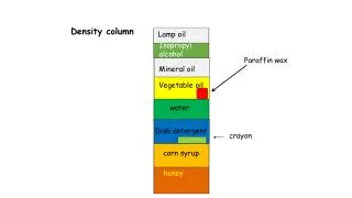
Each disc has • a peripheral part, anulusfibrosus (consisting of concentric layers of collagen fibers), and • a central part the nucleus pulposus (ovoid mass of gelatinous material collagen fibers and a few cartilage cells).
A sudden increase in compression load on vertebral column causes nucleus pulposus to thrust outward, where it may press on the spinal cord or the nerve roots causing pain. • This mainly happens with advancing age collagen fibers of the anulusdegenrates and as a result the anulus cannot contain the nucleus pulposus under stress. • The 1st, 2nd cervical verterbrae, sacrum, and coccyx lacks intervertebral discs
Joints between two vertebral arches • Synocial joints between the supeerior and inferior articular processes of adjacent vertebrae. • The articular facets are covered by hyaline cartilage and sorrounded by a capsular ligament. • Supraspinousligament,interspinous ligament, intertransverse ligaments and ligamentumflavum helps in stabilizing the joints • In cervical region, the supraspinous and interspinous ligaments are greatly thickened to form strong ligamentumnuchae,which extends from 7th cervical spine to the external occipital protuberance
Atlanto-Occipital joints • Synovial joints • Between occipital condyles(present on either side of foramen magnum) and facets on the superior surfaces of the lateral masses of the atlas • Capable of flexion , extension and lateral flexion. They do not rotate
Atlanto-Axial joints • 3 synovial joints; • 1 between odontoid process and the anterior arch of atlas • 2 between the lateral masses of the bones • Extensive rotation of the atlas and thus the head on the axis.
Curves of verterbral column • In fetus, the vertebral column has one continuous posterior convexity. • In adult, the vertebral column exibits • Cervical , posterior concavity • Thoracic, posterior convexity • Lumbar, posterior concavity and • sacral, posterior convexity • In old age, the intervetebral discs atropy • gradual return of vertebral column to continuous posterior convexity