Download
1 / 160
1.68k likes | 2.5k Views
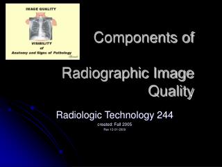
Components of Image Quality & Radiographic Artifacts. Radiologic Technology A Spring 2010 Final. X-ray Exposure Factors Radiographic Density & Contrast Components of Image Quality Radiographic Artifacts. Review Chapter 7. Primary radiation exits the tube

E N D
Components of Image Quality & Radiographic Artifacts Radiologic Technology A Spring 2010 Final
X-ray Exposure Factors • Radiographic Density & Contrast • Components of Image Quality • Radiographic Artifacts
Review Chapter 7 • Primary radiation exits the tube • Interacts with various densities in the body • Photons may be absorbed • Scattered • Passed through without any interference to the cassette or image receptor (IR)
X-ray Exposure Factors • TECHNIQUE SELECTION: • Radiographer selects the • Kilovoltage peak (kVp) • Milliamperage (mA) & time (s) • Milliamperage x time = mAs (milliamperage multiplied by a set time measured in seconds)
Kilovoltage Peak • kVp • One kilovolt = 1000 volts • The amount of voltage selected for the x-ray tube. • Range 30 to 150 kVp • kVp controls __________ ?
Milliamperage • One milliampere (mA) = one thousandth of an ampere. • The amount of current supplied to the x-ray tube • How many x-rays will be produced • Range 10 to 1200 mA
Time • In seconds • How long x-rays will be produced • 0.001 to 6 seconds
Milliampere Seconds • Technologists think in terms of mAs • Calculated by mA x seconds • Ex: 100mA X 0.2s = 20 mAs • How many x-rays will be produced and for how long. • Modern x-ray machines only allow control of • mAs controls _______________ ?
Factors Affecting Density • Primary control factor: • Influencing factors:
Primary Controlling Factor of Density • mAs • mA = AMOUNT of electrons sent across the tube combined with TIME (S) = mAs • mAs controls DENSITY on radiograph primary function of mAs is DENSITY
Imagine this… • If the mA station is changed from 200 to 400 mA, twice as many electrons will flow from the cathode to the anode. • From 10 mA to 1000 mA = 100 x more • mA controls how many electrons are coming at the target • mAs is a combination of how many and for how long(seconds)
10 mA 1000 mA
kVpmore energy = more photons passing though tissue & striking the image • ____________ = doubling of exposure to the film _____________ = halving of exposure to the film _____ rule will also change the contrast of the image because kV is the primary method of changing image contrast. Remember : ___ change ( ) KVP has the same effect as doubling or ½ the MAS on density
Change in kVp • kVp controls the energy level of the electrons and subsequently the energy of the x-ray photons. • A change from 72 kVp will produce x-rays with a lower energy than at 82 kVp • Difference between a ball traveling 72 mph and 82 mph (how much energy did it take to throw the ball at the rates?)
Radiolucent vs. Radiopaque • ___________ materials allow x-ray photons to pass through easily (soft tissue). • __________materials are not easily penetrated by x-rays (bones)
Transmission (no interaction) Responsible for dark areas Scatter (grays) – produces no diagnostic info Absorption (photoelectric effect) Responsible for light areas Creating the Image
Images • ____________ = THE AMOUNT OF BLACKENING “DARKNESS” ON THE RADIOGRAPH (mAs) • ____________ – THE DIFFERENCES BETWEEN THE BLACKS TO THE WHITES (kVp)
Why you see what you see… • The films or images have different levels of density – different shades of gray • X-rays show different features of the body in various shades of gray. • The gray is darkest in those areas that do not absorb X-rays well – and allow it to pass through • The images are lighter in dense areas (like bones) that absorb more of the X-rays.
Image Production • _____________ – The beam of photons, B4 it interacts with the pt’s body. • _____________ – The resulting beam that is able to exit from the patient. • _____________ – Radiation that interacts with matter & only continues in a different direction – not useful for image production. • _____________ – Primary radiation that is changed (partially absorbed) as it travels through the pt.
3 Different Body HabitusHypersthenic Sthenic Hyposthenic Dr. Charman, Eric Guzman, Adam Guzman Thank you to the 3 men in my life ! DCharman
Goal: Producing optimal radiographsDENSITY Too dark Too light
Controlling Factor of Contrast • Kilovolts to anode side – kVp • Kilovolts controls how fast the electrons are sent across the tube • _______ – controls CONTRAST on images
Producing optimal radiographsContrast Scale Long scale short scale
Scale of Contrast? Which one is “better” How does the kVp affect these images?
Creates fog Lowers contrast (more grays) Increases as: Scatter
Collimate to area of interest -reduces scatter and radiation dose to the patient