Preparation of Samples for Scanning Electron Microscopy: A Focus on Radish and Sunflower
This document outlines the preparation methods for plant specimens (Clover, Radish, and Sunflower) intended for viewing under a scanning electron microscope. Key steps include chemical fixation with 5% glutaraldehyde, dehydration through a series of ethanol concentrations, and drying using a critical point dryer with CO2. This process maintains the life-like structure of the samples, preventing deformation. Finally, specimens are coated with a thin layer of graphite to enhance electrical conductivity during imaging, captured at 5 kV using a JEOL JSM-6100 microscope.

Preparation of Samples for Scanning Electron Microscopy: A Focus on Radish and Sunflower
E N D
Presentation Transcript
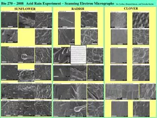
Bio 270 – 2008 Acid Rain Experiment – Scanning Electron Micrographs Liz Casline, Hannah Inman, and Natasha Karim CLOVER RADISH SUNFLOWER How were these samples prepared? To view specimens in an electron microscope, they must be dry, because the instrument is under high vacuum, and they must be electrically conductive, because the electron beam is a current that would otherwise cause heat damage and surface charging. The fresh material was fixed (chemically stabilized) with 5% glutaraldehyde (in pH 7.2 phosphate buffer) at 4 C for 12 hours. The samples were then dehydrated by running them through an alcohol series (25, 50, 75, 95, 100, 100, 100% ethanol) for 4-12 hours per step. The samples were then dried using a critical point dryer using CO2. The samples are soaked in liquid carbon dioxide to flush the ethanol from the tissues, and then are brought to the critical point of carbon dioxide (where the liquid and gas phases exist in equilibrium – 42 C and 1300 PSI.) By slowly releasing the pressure while maintaining the critical temperature, the CO2 leaves the tissues without causing any deformation, that is, the dry tissue retains its life-like structure. For example, a grape prepared this way would still look like a grape at the end. An air-dried grape would become a raisin due to surface-tension effects. The dried specimens are then coated with a thin (20 nm) film of graphite (carbon) using a device called a carbon evaporator. The coating makes the specimens electrically conductive so that they won’t be damaged by the electron beam of the microscope. The specimens were viewed at 5 kV using a JEOL JSM-6100 scanning electron microscope. The images were captured using AIA2 Digital Imaging Systems software.