Download
1 / 18
180 likes | 281 Views
LGN, superior colliculus, and optic tectum development and innervation. Chris Strang Ph.D. Vision Sciences WORB 308 cstrang@uab.edu 975-7222. Learning Objectives. LGN specification Zona limitans- Shh Timing of LGN lamination Superior colliculus and tectum specification Isthmus

E N D
LGN, superior colliculus, and optic tectum development and innervation Chris Strang Ph.D. Vision Sciences WORB 308 cstrang@uab.edu 975-7222
Learning Objectives • LGN specification • Zona limitans- Shh • Timing of LGN lamination • Superior colliculus and tectum specification • Isthmus • FGF8 and transcription factors Pax2, En1, and Otx2 • Timing of colliculus lamination • Target innervation- gradients of ligands; receptors • Ephrin-A2, Ephrin-A5; ephA3 • Ephrin-B; ephB • Wnt3; Ryk • EphrinB1; ephB2, ephB3
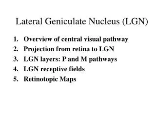
Once axons extending from ganglion cells have found their way to the targets they must invade the target and find the proper location either in the LGN and superior colliculus (mammals), or optic tectum (non-mammals). The axons must maintain retinotopic mapping, and segregate into eye specific and cell-type specific layers. But, first, where and how do these targets arise?
The LGN of the thalamus is the main target of retinal projections in mammals. The thalamus arises in the diencephalon. It is induced by Shh from the zona limitans, the organizing region at the p2-p3 border. The zona limitans becomes the boundary between the prethalamus and thalamus. LGN cells are born at the wall of the third ventricle at the midline and migrate to positions at the lateral margin of the thalamus, and are then arranged in layers.
At the time that the retinal afferents arrive, the LGN is incompletely laminated. Glial cells are distributed in layers after retinal afferents arrive but before LGN neurons show lamination. The adult pattern of cell rich layers interleaved with cell free layers arises after innervation and glial redistribution.
The colliculus and tectum are analogous midbrain structures. In mammals, the superior colliculus is involved in eye movements, including saccades. It also receives and integrates non-visual inputs. It is homologous to the non-mammalian optic tectum, but the tectum, rather than the LGN, is major the visual path in non-mammals. For mammals and non-mammals, the posterior midbrain boundary is defined by expression patterns of Wnt1, Otx2, Gbx2, and FGF8 at the isthmus. Otx2 (pink) is expressed from the forebrain to the isthmus at the midbrain-hindbrain border. Fgf8 (orange) is expressed at the isthmus. Gbx2 (blue) is expressed in the anterior hindbrain region. En1, En2 and Pax2 (hatched red) are expressed in the midbrain and in the anterior hindbrain. Wnt1 (purple) is expressed at the posterior midbrain and at the dorsal midline.
Pax2, En1, and FGF8 interact in specification of the tectum. Overexpression of any of these factors at the level of the diencephalon results in ectopic tectum formation. Interestingly, overexpression of Otx2 in the hindbrain also results in ectopic tectum formation, indicating that Otx2 is also a required factor. Shh may be instrumental in limiting tectum development to the dorsal neural tube. Overexpression of Shh on one side results in elimination of tectum on that side. Nakamura, (2001) TINS, 24:32-39
Engrailed 1 and 2 are also important in retinal targeting to superior colliculus. The En gradients are high at the isthmus, and decrease further anteriorly (En1) and posteriorly (En2). Reversal of the isthmus can reverse engrailed gradients and subsequent axon innervation.
Lamination and innervation of the superior colliculus. In humans at embryonic week 8, the presumptive colliculus consists of one layer of proliferating progenitor cells at the ventricular zone. Cell rich areas begin to appear around embryonic week 11. At approximately 14 weeks fiber layers begin to appear. The characteristic alternation of white and grey matter layers seen in the mature superior colliculus is evident by embryonic week 16. Retinal axons begin to invade the superficial layers by week 12, and penetrate into the deeper layers by week 13. Qu et al, (2006) Experimental Eye Research, 82:300-310
However, while prenatal retinal inputs have adult-like organization, the SC cells themselves are functionally immature until sometime after birth. Studies in cat have demonstrated that maturity of the superior colliculus projections to upstream brain areas may develop from center to periphery. For example, the ability to orient to visual cues is dependent on superior colliculus, and the ability to orient to cues in the center of the visual field develops before the orienting response to cues in the periphery. Qu et al, (2006) Experimental Eye Research, 82:300-310
Retinotopic mapping must be maintained when axons invade the target to form synaptic connections. Studies in the frog first gave solid evidence for the possibility that there are specific molecular cues that determine how axons invade the target tissue. How do axons from anterior retina choose posterior tectum, and axons from the posterior retina choose anterior optic tectum?
A series of crucial experiments showed that there are specific molecules in the tectum itself that inhibit axon growth. Concentration varied with tectal position, and the sensitivity of retinal axons to the factor varied with retinal position. These factors turned out to be the receptor tyrosine kinases called the eph kinases, and their membrane associated ligands, the ephrins.
These studies lead to the identification of 2 RTK ligands, ephrin-A2 and ephrin-A5. These are expressed in low to high gradients in the anterior direction in the tectum. The complimentary Eph receptors are expressed by chick retinal ganglion cells in a low to high nasal to temporal gradient. These receptors, ephA3, bind to both ephrin-A2 and A5. The ephrins inhibit temporal retina axon growth so the axons stop in the anterior tectum. Axons from anterior (nasal) retina express less ephA3 and so can continue to grow into posterior tectum. There is a second gradient of Wnt3 which is highest in medial tectum and lowest laterally. The Wnt receptor is highest in ventral retina. Thus each axon has a “preference” for specific locale in tectum. This preference is probably manifested by a difference in adhesion, with signaling making the substrate more or less permissive for axon growth.
As in frog tectum, GCs in mouse nasal retina express low EphA and project to posterior superior colliculus, which has a high concentration of ephrin-A. GCs in temporal retina have high expression of EphA and project to anterior colliculus. Dorsal and ventral GC mapping to lateral and medial SC is mediated by an ephrin-B gradient and EphB receptors. Low expression of EphB receptors by dorsal GCs maps to the lateral (ventral) SC. GCs in ventral retina have high expression of EphB and project to the medial (dorsal) SC. Topographical organization of retinogeniculate projections are also specified by ephrin gradients. GCs in nasal retina project to dorsal contralateral LGN. GCs in temporal retina project to ipsilateral ventral LGN. RGCs in ventral retina project to the medial LGN, while GCs in dorsal retina project to lateral LGN.
Wnt3 is expressed in a medial to lateral gradient in chick tectum and mouse colliculus. The Wnt3 receptor, Ryk, is expressed by retinal ganglion cells in a gradient that decreases from ventral to dorsal in GCs. GC axon termination zones are repelled by Wnt3 expression. Wnt3 activation of different receptor, Frizzled, mediates attraction. These cues interact with the gradient established by Eph receptors to provide a mechanism for mapping and axon branching in the tectum. Schmitt et al., (2006) Nature 493(5):31-36
Wnt3 signaling mediated by Ryk appears to provide for lateral mapping. Ventral axons are repelled by Wnt3, while dorsal axons are attracted by low Wnt3 and repelled by high Wnt3. EphrinB1 activation of EphB2 and EphB3 receptors provides medial mapping by attracting axon branches. When Wnt3 repulsion is blocked, ephrinB1–EphB signaling causes branches to project medially. When ephrinB1–EphB signaling is eliminated, interstitial branches are repelled from the medial side by Wnt3 and project laterally.
In many species, axon innervation of the target must be refined. As in humans, the segregation of the cat colliculus into eye specific bands takes place prior to birth. Direct retinal input is initially only to the superficial layers and axons from both eyes invade along the entire colliculus. The ipsilateral projection retracts later. In the LGN of cats and primates, GCs project to eye-specific LGN layers (LGN projects to layer IV of visual cortex forming ocular dominance columns). However, althought GC axons projecting to the LGN invade the correct target region, they initally extend a little beyond and then pull back. It turns out that the axons actually make two kinds of wrong projections. Prenatal axons branch in the wrong layer and the terminal arborization in the correct layer is too wide. Improper connections in the LGN and SC are pruned back postnatally. Axon innervation and map refinement are activity dependent but begin before visual experience.
Learning Objectives • LGN specification • Zona limitans- Shh • Timing of LGN innervation and lamination • Superior colliculus and tectum specification • Isthmus • FGF8 and transcription factors Pax2, En1, and Otx2 • Timing of colliculus innervation and lamination • Retinotopic mapping- gradients of ligands; receptors • Ephrin-A2, Ephrin-A5; ephA3 • Ephrin-B; ephB • Wnt3; Ryk • EphrinB1; ephB2, ephB3