Download
1 / 1
10 likes | 209 Views
No. 030. Peptide -Based Identification of Exosome Proteins from Prostate Cancer Cell Lines . Vicki M Velonas*, Mark Raftery ^, Ling Zhong ^, Anne Poljak ^ ∞ , Stephen Assinder *, Henry Woo # , and Cris dos Remedios* *Bosch Institute, The University of Sydney

E N D
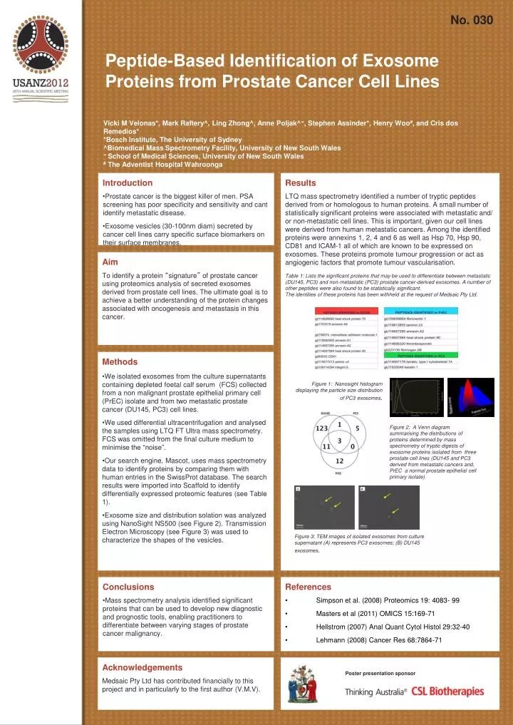
No. 030 Peptide-Based Identification of Exosome Proteins from Prostate Cancer Cell Lines Vicki M Velonas*, Mark Raftery^, Ling Zhong^, Anne Poljak^∞, Stephen Assinder*, Henry Woo#, and Cris dos Remedios* *Bosch Institute, The University of Sydney ^Biomedical Mass Spectrometry Facility, University of New South Wales ∞ School of Medical Sciences, University of New South Wales # The Adventist Hospital Wahroonga • Introduction • Prostate cancer is the biggest killer of men. PSA screening has poor specificity and sensitivity and cant identify metastatic disease. • Exosome vesicles (30-100nm diam) secreted by cancer cell lines carry specific surface biomarkers on their surface membranes. Results LTQ mass spectrometry identified a number of tryptic peptides derived from or homologous to human proteins. A small number of statistically significant proteins were associated with metastatic and/ or non-metastatic cell lines. This is important, given our cell lines were derived from human metastatic cancers. Among the identified proteins were annexins 1, 2, 4 and 6 as well as Hsp 70, Hsp 90, CD81 and ICAM-1all of which are known to be expressed on exosomes. These proteins promote tumour progression or act as angiogenic factors that promote tumour vascularisation. Table 1: Lists the significant proteins that may be used to differentiate between metastatic (DU145, PC3) and non-metastatic (PC3) prostate cancer-derived exosomes. A number of other peptides were also found to be statistically significant. The identities of these proteins has been withheld at the request of Medsaic Pty Ltd. Aim To identify a protein “signature” of prostate cancer using proteomics analysis of secreted exosomes derived from prostate cell lines. The ultimate goal is to achieve a better understanding of the protein changes associated with oncogenesis and metastasis in this cancer. • Methods • We isolated exosomes from the culture supernatants containing depleted foetal calf serum (FCS) collected from a non malignant prostate epithelial primary cell (PrEC) isolate and from two metastatic prostate cancer (DU145, PC3) cell lines. • We used differential ultracentrifugation and analysed the samples using LTQ FT Ultra mass spectrometry. FCS was omitted from the final culture medium to minimise the “noise”. • Our search engine, Mascot, uses mass spectrometry data to identify proteins by comparing them with human entries in the SwissProt database. The search results were imported into Scaffold to identify differentially expressed proteomic features (see Table 1). • Exosomesize and distribution solation was analyzed using NanoSightNS500 (see Figure 2). Transmission Electron Microscopy (see Figure 3) was used to characterize the shapes of the vesicles. Figure 1: Nanosight histogram displaying the particle size distribution of PC3 exosomes. Figure 2: A Venn diagram summarising the distributions of proteins determined by mass spectrometry of tryptic digests of exosome proteins isolated from three prostate cell lines (DU145 and PC3 derived from metastatic cancers and, PrEC a normal prostate epithelial cell primary isolate). Figure 3: TEM images of isolated exosomes from culture supernatant (A) represents PC3 exosomes; (B) DU145 exosomes. • Conclusions • Mass spectrometry analysis identified significant proteins that can be used to develop new diagnostic and prognostic tools, enabling practitioners to differentiate between varying stages of prostate cancer malignancy. • References • Simpson et al. (2008) Proteomics 19: 4083- 99 • Masters et al (2011) OMICS 15:169-71 • Hellstrom (2007) Anal Quant CytolHistol 29:32-40 • Lehmann (2008) Cancer Res 68:7864-71 Acknowledgements Medsaic Pty Ltd has contributed financially to this project and in particularly to the first author (V.M.V). Poster presentation sponsor DU145 PC3 DU145 PC3 PrEC PrEC A B 100nm 100nm