Download
1 / 20
210 likes | 331 Views
This case study examines a rare phacoantigenic response to a ruptured lens capsule, showcasing a granulomatous reaction and treatment options. Phacoantigenic response occurs post-surgery or trauma, sensitizing the individual to their own lens protein. Treatment involves corticosteroids and aqueous suppressants.

E N D
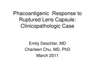
Phacoantigenic Response to Ruptured Lens Capsule: Clinicopathologic Case Emily Deschler, MD Charleen Chu, MD, PhD March 2011
Brief History and Course • 58 year-old woman • Blind left eye since teens – unknown etiology • No history of trauma • Referred to glaucoma service when left eye became painful
Vision: Right eye: 20/20 Left eye: NLP IOP: Right eye: 11 Left eye: 16 Pupil: Right eye: 4-3 Left eye: Irregular Anterior segment Right eye: Shallow anterior chamber with patent PI Left eye: injected conjunctiva, cornea with PEE, shallow anterior chamber with patent PI x2, iris with florid rubeosis, posterior synechiae, mature cataract Fundus: Right eye: Normal, c/d 0.6 with healthy neuroretinal rim Left eye: no view Initial Physical Exam
Brief History and Course • Symptoms initially attributed to ocular surface disease and treated with artificial tears, topical steroids • 1 year later, pain continued and patient elected to proceed with enucleation (PHS10-35123)
Calcific Band Keratopathy • Gross photo of the cornea • entire cornea is affected • calcification spans from limbus to limbus
Calcific Band Keratopathy • Seen clinically as calcific plaques in the interpalpebral zone • deposition of calcium in the epithelial basement membrane, Bowman’s layer, and anterior stroma. • usually an intervening region of cornea between the limbus and the calcification that is unaffected • 6 main causes • chronic ocular disease (usually inflammatory), hypercalcemia, hereditary transmission, elevated serum phosphorus with normal calcium, exposure to mercury, silicone oil in ahakic eye • Tx: remove overlying epithelium and then apply EDTA
H&E 1. stippled basophilia of Bowman’s layer, the calcium deposits merge to form a linear array along Bowman’s layer 2. keratocyte nuclei Calcific Band Keratopathy 2 1
Neovascularization of the Iris & Hyphema 1. Neovascularization of the iris – at this power cannot see individual vessels, but flattened anterior iris is a clue 2. Hyphema 1 2
Systemic vasular disease Carotid disease Giant cell arteritis Ocular vascular disease Diabetes CRVO Coats Eales ROP Persistent fetal vasculature Anterior segment ischemia Ocular disease Cronic uveitis Chronic retinal detachment Endophthalmitis Stickler syndrome Retinoschisis Intraocular tumors Uveal melanoma Metastatic disease Retinoblastoma Ocular therapy Radition therapy Disorders Predisposing to Neovascularization of Iris and Angle
Peripheral Anterior Synechia • Iris stroma obliterates the angle recess • Mechanisms of PAS formation: • Contraction of an inflammatory, hemorrhagic or vascular membrane, band or exudate in the angle • Forward displacement of the iris-lens diaphragm leading to a prolonged shallow/flat anterior chamber • Extent of PAS may correlate with visual field damage, larger vertical cup-to-disc ratio, and higher untreated intraocular pressure • Poor prognosis • Treat underlying cause (ischemic retina with PRP) anti-VEGF, filtering surgery
1. Chronic Iritis 2. Neovascularization of the Iris • Lymphocytes • small cells with round, hyperchromatic nuclei and scant cytoplasm • found in chronic inflammatory process 1 2
Chronic Plasmacytic Cycloiritis • B lymphocytes may differentiate into plasma cells and produce immunoglobulin • Eccentric “cartwheel” or “clockface” nuclei
Chronic Choroiditis • Small blue cells
Phacoantigenic Response to Ruptured Lens Capsule • Granulomatous reaction • Macrophages filled with degenerated lens cortical material
Phacoantigenic Sensitized to own lens protein following surgery or penetrating trauma Granulomatous reaction Clinically Moderate anterior chamber reaction with KP on endothelium and lens capsule Low grade vitritis Synechial formation Not commonly associated with glaucomatous optic neuropathy Treatment Corticosteroids, aqueous suppressants, and if unsuccessful, remove residual lens material Phacolytic Leakage of lens proteins through the capsule of a mature or hypermature cataract Proteins, phagocytizing, inflammatory debris obstruct the uveal meshwork Clinically Prominent cell and flare with possible psuedohypopyon in the anterior chamber No KP Morgagnian cataract with wrinkled anterior lens capsule Open anterior chamber angle Treatment IOP lowering medications, ultimately cataract extraction Phacoantigenic vs PhacoanalyticGlaucoma
Discussion: Phacoantigenic response to ruptured lens capsule • Occurs following surgery or penetrating trauma causing sensitization to one’s own lens protein • Granulomatous reaction • Variable clinical presentation: moderate AC reaction with KP on corneal endothelium and anterior lens surface (unlike phacolytic) with moderate AC reaction. Low grade vitritis, synechial formation, residual lens material may be in AC • Not commonly associated with glaucomatous optic neuropathy. • Treatment • Corticosteroids, aqueous suppressants, and if unsuccessful, remove residual lens material
Discussion: Phacolytic response to mature cataract • Proteins, phagocytizing macrophages, and other inflammatory debris obstruct the trabecular meshwork • Generally an elderly person with a history of poor vision presents with sudden onset pain, conjunctival injection, elevated IOP, corneal edema, prominent cell and flair without KP and open anterior chamber angle • Cellular debris may be present in the anterior chamber presenting as pseudohypopyon. The anterior capsule may appear wrinkled representing loss of lens material • Treatment: • IOP lowering medications, ultimately cataract extraction
Diagnosis: • Phacoantigenic immune response to ruptured lens capsule • Additional diagnoses: • Idiopathic chronic panophthalmitis • Neovascularization of the iris • Peripheral anterior synechiae • Calcific band keratopathy
Sources • Basic Clinical Science Course Section 10: Glaucoma. American Academy of Ophthalmology 2009-2010 • Yanoff, Myron and Fine, Ben. Ocular Pathology. Mosby. 2002