Lupus neprhitis
Lupus neprhitis. Staci Smith DO Nephrology Grandview Hospital. Today’s objectives. Overview of Lupus Types of lupus History Common manifestations SLE Nephritis WHO classification Biopsy Indications Biopsy Findings Treatment. Differential Diagnosis. hematuria


Lupus neprhitis
E N D
Presentation Transcript
Lupus neprhitis Staci Smith DO Nephrology Grandview Hospital
Today’s objectives • Overview of Lupus • Types of lupus • History • Common manifestations • SLE Nephritis • WHO classification • Biopsy Indications • Biopsy Findings • Treatment
Differential Diagnosis • hematuria • proteinuriaglomerulonephritis • red blood cell casts
SLE Minimal Change Dz Membranous GN FSGS MPGN RPGN Ig A Nephropathy Anti GBM Dz Goodpasture’s Wegener’s Hepatitis B, C AIDS Amyloidosis HSP Cryoglobulinemia Vasculitides Poststrept/ Poststaph GN DDx : GlomerulonephriticDz
Red Blood Cell Casts • red cell casts • virtually diagnostic of glomerulonephritis or vasculitis • only one needed • absence does not exclude diagnosis
Types Of Lupus • Systemic Lupus: • most common and affects major organs • Discoid Lupus: • affects only the skin • not fatal, but can cause severe scarring • Drug-induced Lupus: • is systemic Lupus caused by medications • when the medicine is stopped, the disease goes away
What is Systemic Lupus Erythematous? • autoimmune disorder • multisystem microvascular inflammation • defined by clinical picture and generation of autoantibodies • mostly against double stranded DNA
Pathogenesis of SLE • autoantibodies • mostly against double stranded DNA and the Smith antigen • Ab to Smith (Sm) antigen is very specific for SLE • 25% of patients
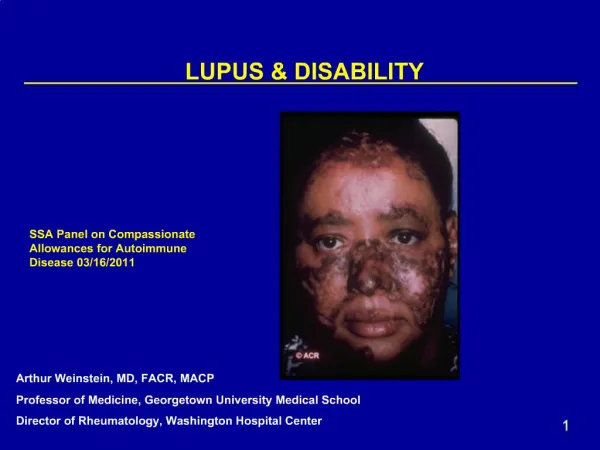
History of SLE • not known when Lupus first appeared • Hippocrates noted similar diseases in Ancient Greece • facial rash that resembles the markings of a wolf • 1851 French-man named Pierre Cazenave • first clinical records • more than 1.4 million Americans are affected by SLE
ANA Antibodies Rim Diffuse Speckled Nucleolar
Lupus Criteria • American College of Rheumatology • presence of 4 of 11 criteria can establish SLE Dx • 96% sensitive and specific • updated 1995
Serositis –pleuritis, pericarditis Oral ulcers - painless Arthritis – 2 or more peripheral joints Photosensitivity Blood Abnormalities –thrombocytopenia, lymphopenia, lymphopenia (x2),hemolytic anemia Renal – casts, proteinuria, hematuria ANA positive Immune Abnormalities – ANA, Anti DS DNA, Smith Ag, false (+) syphilis Neurologic - seizures, psychosis Malar Rash- spares nasolabial folds Discoid Rash – scaling,scaring American College of Rheumatology Criteria for Diagnosis of SLE SOAP BRAIN MD
Lupus and the Kidney • Lupus nephritis • one of the most serious manifestations of SLE • typically arises within 5 years of diagnosis • commonly within the first 6 to 36 months • Renal failure rarely occurs before American College of Rheumatology classification criteria are met.
Lupus and the Kidney • total incidence of renal involvement among patients with SLE exceeds 90 % • abnormal urinalysis • with or without an elevated Cr • in approximately 50% at diagnosis time • proteinuria present in 80% • 40% have hematuria and/or pyuria
Lupus and the Kidney • ‘Silent’ lupus nephritis • normal urinalysis • no proteinuria • normal serum creatinine levels • However, renal biopsy reveals pathological changes
Lupus Nephritis • Six types of renal involvement with SLE • Why do renal biopsy? • to determine stage of disease • histological evidence is present in most SLE pts even if they do not have clinical manifestations of renal disease • Pattern of glomerular injury • related to the site of formation of the immune deposits • is primarily due to anti DS DNA
Morphological Classification of Lupus Nephritis (modified WHO Classification)
Normal Glomerulus • light micrograph • capillary lumens open • glomerular capillary wall thickness • similar to that of the tubular basement membranes • mesangial cells and matrix are located in the central or stalk regions of the tuft
Mesangial Proliferative Lupus Nephritis: Class II • segmental areas of increased mesangial matrix and cellularity • light micrograph
Focal Proliferative Nephritis (Class III) Subsets • Divided by active and/or chronic lesions: • Class III (A): • active lesions • Class III (A/C): • active and chronic pathology • Class III (C): • chronic inactive lesions with scarring • a.k.a. focal sclerosing lupus nephritis
Focal Proliferative Nephritis (Class III) • usually associated with subendothelial deposits • areas of cellular proliferation • thickening of glomerular capillary • “wire loop”
Diffuse Proliferative NephritisClass IV • subendothelial deposits • deposition of immunoglobulins and complement • results in thickening of the glomerular capillary wall • subsets • segmental = < 50% of glomeruli • diffuse = >50% of glomeruli
Diffuse Proliferative Nephritis:Class IV • subendothelial deposits • thickening of glomerular capillary wall
Membranous Nephritis • Class five • the one form of lupus nephritis that may present with no other clinical or serologic manifestations of SLE • typically presents with signs of nephrotic syndrome • microscopic hematuria and hypertension also may be seen • Cr concentration is usually normal or only slightly elevated
Sclerosing Nephritis :Class VI • sclerosis of more than 90% of glomeruli • represents healing of previous inflammatory injury • as well as the advanced stage of chronic class III, IV, or V lupus nephritis • immunosuppressive therapy is NOT likely to be beneficial
diffuse (class IV) or severe focal (class III) proliferative glomerulonephritis, • severe or progressive membranous lupus (class V) • marked nephrotic syndrome • rising serum creatinine • membranous in association with class III or class IV disease • mixed disease
Therapy for lupus patients with arthritis • No internal organ involvement • First line: NSAID’s • Cyclooxygenase-2 specific inhibitor • may induce thrombotic risk in patients with antiphospholipid antibodies • Low dose hydroxychloroquine • 200mg twice a day
Manifestations not often responsive to glucocorticoids • Thrombosis—includes strokes • Glomerulonephritis • Resistant thrombocytopenia or hemolytic anemia
Therapy for patients with lupus nephritis • Previously untreated patients • Active lupus nephritis or severe manifestations • decreased renal function and /or high-grade proteinuria • First line: high doses of corticosteroids • about 1mg/kg/day • Cytotoxic drugs or other immunosuppressive drugs
The indications of cytotoxic drugs use in the treatment of lupus nephritis • Active and severe GN depsit high dose steroids • Responded to corticosteroids but require an unacceptably high dose to maintain a response. • Side effects from corticosteroids • Chronic damage on a renal biopsy
Use of Cytotoxic Drugs in SLE : Azathioprine • requires 6–12 months to work well • 1–3 mg/kg/day(initial dose) • 1–2 mg/kg/day(maintenance dose) • Advantage:probably reduces flares, reduces renal scarring, reduces glucocorticoid dose requirement • Side effects: Bone marrow suppression, leukopenia, infection(herpes zoster), infertility, malignancy, early menopause, hepatic damage, nausea
Advantage • reduces flares, reduces renal scarring, reduces glucocorticoid doses • Side effects • bone marrow suppression, leukopenia, infection, malignancy, nausea,etc
Use of Cytotoxic Drugs in SLE: Cyclophosphamide • requires 2–16 weeks to work well • Initial dose:1-3 mg/kg/day orally or 8–20 mg/kg intravenously once a month plus mesna • Maintenance dose:0.5–2 mg/kg/day orally or 8–20mg/kg intravenously every 4–12 wks • Mesna
mycophenoalte mofetil may be an alternative to cyclophosphamide as initial therapy • particularly among patients who refuse or cannot tolerate cyclophosphamide • Biggest side effect is diarrhea, also myelosuppression • fewer side effects than cyclophosphamide
Rituximab • interferes with the activation and differentiation of B cells • lysis mediated by: • Complement • Fc receptor-bearing cytotoxic cell • Inducing apoptosis • selective transient depletion of the CD20+ B-cell subpopulation
Other management principles in the treatment of lupus patients • Avoid possible disease triggers-sulfa antibiotics, sun, high estrogen-containing birth control pills,alfalfa sprouts • Prevent atherosclerosis • Prevent osteoporosis • Prevent infection • Prevent progression of renal disease • Prevent clots in patients with antiphospholipid antibodies
Differential Diagnosis hematuria proteinuriaglomerulonephritis red blood cell casts
What is Systemic Lupus Erythematous? autoimmune disorder multisystem microvascular inflammation defined by clinical picture and generation of autoantibodies mostly against double stranded DNA
American College of Rheumatology Criteria for Diagnosis of SLE Serositis –pleuritis, pericarditis Oral ulcers - painless Arthritis – 2 or more peripheral joints Photosensitivity Blood Abnormalities –thrombocytopenia, lymphopenia, lymphopenia (x2),hemolytic anemia Renal – casts, proteinuria, hematuria ANA positive Immune Abnormalities – ANA, Anti DS DNA, Smith Ag, false (+) syphilis Neurologic - seizures, psychosis Malar Rash- spares nasolabial folds Discoid Rash – scaling,scaring SOAP BRAIN MD
Morphological Classification of Lupus Nephritis (modified WHO Classification)