Download
1 / 25
260 likes | 475 Views
Learn to assess patients with thyroid nodules, determine surgical needs, and diagnose pathologies. Understand thyroid function tests and interpretation of FNA results. Identify indications for surgery and the workup process.

E N D
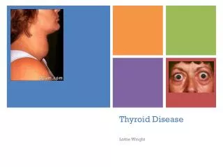
Thyroid Disease Nodules and Neoplasms By: Christine B. Taylor, MD
Learning Objectives After reviewing this module, the student will have the ability to: • Approach History & physical exam of patients with thyroid pathology • Understand which patients require surgery and appropriate surgical timing • Describe the process of evaluating and working up various thyroid pathologies
Case Presentation • 29 year old Female referred to Head & Neck clinic for evaluation of a thyroid nodule. Patient reports this nodule was found incidentally while she was getting ready for work one morning. • She went to her PCP, who ordered a thyroid ultrasound, which demonstrated a 2cm nodule in the right lobe of the thyroid.
Question • After thorough history and physical, what would you order first for this patient? • A. Thyroid function tests (TSH, T4) • B. CT neck • C. MRI neck • D. Radioactive Iodine uptake scan • E. All of the above
Question • After thorough history and physical, what would you order first for this patient? • A. Thyroid function tests (TSH, T4) • B. CT neck • C. MRI neck • D. Radioactive Iodine uptake scan • E. All of the above
Explanation • What would you order first for this patient? • A. Thyroid function tests (TSH, T4) It is important to first establish the patient’s thyroid function. This will help determine if the known thyroid nodule is hyperfunctioning, hypofunctioning, or nonfunctioning. At this point, you should also obtain Fine Needle Aspiration (FNA) with ultrasound guidance, if necessary, to obtain cells for cytopathology. This will help determine if nodule is benign or malignant
Important Points - Patient History • Family history of thyroid disease or thyroid cancer? • Familial syndromes (MEN) • Personal history of radiation to head/neck • Increased risk of thyroid cancer • Hoarseness, SOB, difficulty swallowing • Compressive symptoms of thyroid goiter
Case Presentation • Patient reports her voice seems to have become slightly more “husky” lately. She recalls only occasional discomfort with sensation that something is “pushing” in. Denies shortness of breath. • Denies family history of thyroid cancer • Denies personal history of radiation therapy or thyroid or any other type of cancer
Physical Exam • What components of the physical exam are critical for this patient? • Full Head and neck exam to look for any “lumps or bumps” • Palpate for lymphadenopathy • Palpate thyroid for nodularity, firmness or hard masses • Fiberoptic or direct laryngoscopy to evaluate vocal cord function
Case Presentation • On physical exam, you palpate a grossly enlarged thyroid gland with a 1.5cm dominant nodule on the right thyroid lobe • You discover the following findings on fiberoptic exam: View on laryngoscopy Upon inspiration: Symmetric bilateral Vocal fold abduction Opening of esophagus trachea Vocal folds (true cords) Epiglottis False vocal folds Base of tongue/lingual tonsil anterior
Question • Patient is sent for labs as well as FNA. Patient returns to clinic the following week with FNA report reading “follicular cells, cannot rule out follicular neoplasm”. Is surgery indicated for your patient at this time? • Yes • No
Question • Patient is sent for labs as well as FNA. Patient returns to clinic the following week with FNA report reading “follicular cells, cannot rule out follicular neoplasm”. Is surgery indicated for your patient at this time? • Yes • No
Explanation • Surgery is indicated. Follicular cells on FNA can be a benign finding or may indicate follicular carcinoma. • Follicular carcinoma cannot be diagnosed solely on FNA (normal thyroid contains follicular cells.) • Perform at least hemithyroidectomy for tissue diagnosis • Tissue taken at time of surgery must be sent for pathology to evaluate for extracapsular extension, lymphovascular invasion, or metastasis. • Therefore, in the case that follicular neoplasm is suspected based on H&P and FNA results, patient should be taken to surgery for pathologic diagnosis and treatment.
Indications for Thyroidectomy • Hyperthyroidism (Grave’s) not responsive to medical therapy with ophthalmic symptoms • Malignancy (confirmed or high suspicion based on history and/or FNA) • Goiter with compressive symptoms • Large thyroid nodule (>2cm) that is unable to be adequately sampled by FNA (sampling error due to large area of nodule and risk of combination of benign and malignant cells)
Etiologies of Thyroid nodules • Malignant • Papillary carcinoma • Follicular carcinoma • Hurthle cell tumor • Medullary Thyroid Carcinoma • Anaplastic Carcinoma • Lymphoma of thyroid • Benign • Benign thyroid cysts (degenerated nodules) • Colloid nodules • Multinodular goiter
Tests • - CBC, Chemistry • Chemistry for calcium, to include albumin to calculate corrected calcium • Corrected Ca = 0.8 x (4 - patient albumin) + ptCa • - Thyroid function (TSH, free T4) • - Parathyroid hormone (PTH) • - Coagulation studies (INR, PTT) • - FNA of nodule (+/- Ultrasound guidance) • - CT Neck for surgical planning, if appropriate
Imaging • - Ultrasound • - CT Neck for surgical planning CT scan demonstrating large Right thyroid mass causing tracheal deviation to left
Thyroid ULTRASOUND Normal thyroid and surrounding neck anatomy
Treatment • Medical Management • Involve endocrinology early to assist in management • Thyroid hormone replacement (Levothyroxine) for hypothyroidism • Thyroid suppression for hyperthyroidism • I-131 for medical thyroid ablation • Observation for benign nodules • Surgery • Failure of medical management • Malignancy or concern for malignant potential • Symptom management
Treatment • - Post-surgical therapy • I-131 : Radioactive iodine ablation may be indicated postoperatively for any residual malignancy • Thyroid hormone replacement after total thyroidectomy • Calcium replacement • Surgery to thyroid/parathyroid bed • - Ensure patient has endocrinology follow up • Titrate levothyroxine and help manage long term iatrogenic hypothyroidism
Algorithm/Overview Thyroid nodule/mass FNA indeterminate Benign Repeat FNA Malignant or suspicious Follow clinically Tissue pathology surgery
Complications of thyroidectomy • Intraoperative • Bleeding • Damage to arteries/veins of neck • Postoperative presentation • Injury to recurrent laryngeal nerve • Unilateral: hoarseness • Bilateral: respiratory distress • Bleeding • Expanding hematoma – causes compression, shortness of breath • Hypocalcemia • Removal or injury to parathyroid glands or their blood supply • Scar If patient develops expanding neck hematoma postoperatively, treatment involves immediate opening of sutures to evacuate clot and return to OR to explore and stop bleed
Take home Points • - Always obtain thyroid function tests as part of intial workup in patient with thyroid pathology • - Perform full Head & Neck exam • - Thyroid nodules should undergo FNA for cytopathology results
Resources • Flint. Cummings Otolaryngology: Head & Neck Surgery, 5th ed. 2010 Mosby/Elsevier. • Kwak, et al. Findings of Extrathyroid Lesions Encountered With Thyroid Sonography. J Ultrasound Med 2007; 26:1747–1759 • Netter Atlas of Anatomy – Accessed via The Netter presenter : human anatomy collection. edited by John T. Hansen. Elsevier 2010. • Pasha, Raza. Otolaryngology Head & Neck Surgery: Clinical Reference Guide. 3rd edition • Shahi et al. Journal of Medical Case Reports 2010 4:15 doi:10.1186/1752-1947-4-15 • Photo credit: UT Voice Center Website http://uthscsa.edu/oto/voicecenterhome.asp
Acknowledgements • Thank you to Christian Stallworth, MD for overseeing this project and providing advisory support. • Photo credits- Thank you to Megan M. Gaffey, MD and Lauren Thomas for providing photos of the physical exam • Editing and input: