Download

1 / 20
200 likes | 506 Views
Carotid Imaging Modalities. Kyle Boyce August 3, 2007. Leading Causes of Death in U.S. 1. Heart Disease 2. Cancer 3. Stroke (2 nd leading cause Worldwide) Carotid Artery Atherosclerosis – 7% of patients presenting with initial stroke. Non-modifiable Age Race Gender Family History

E N D
Carotid Imaging Modalities Kyle Boyce August 3, 2007
Leading Causes of Death in U.S. • 1. Heart Disease • 2. Cancer • 3. Stroke (2nd leading cause Worldwide) • Carotid Artery Atherosclerosis – 7% of patients presenting with initial stroke
Non-modifiable Age Race Gender Family History Genetics Modifiable *HTN* Hyperlipidemia Diabetes Smoking EtOH High Homocysteine Low Folate Risk Factors
Brief Review - Pathophysiology • Fatty streaks • Intimal thickening • Fibrous Plaque • Increased Smooth muscle cells • Accumulation of connective tissue • Lipid pool • Advanced lesions • Re-vascularized • Necrotic lipid-rich core
Cerebrovascular Symptoms • Ipsilateral Partial or Complete Blindness • Absent Pupillary Light Response • Contralateral Hemianopsia • Contralateral Hemiparesis • Contralateral Sensory Loss • Aphasia (Left Hemisphere Ischemia) • Left visuospatial Neglect (Right Hemisphere) • Atypical findings include Limb shaking and Syncope (not generally considered a result of carotid stenosis)
Measuring Degree of Stenosis Currently, three methods (NASCET, ECST and CC) predominate worldwide.
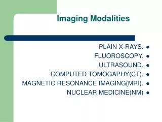
Imaging • Carotid Duplex Ultrasound • CT angiography • MR angiography • Cerebral Angiography (gold standard)
Carotid Duplex U/S • Uses B-mode ultrasound imaging and Doppler ultrasound to detect focal increases in blood flow velocity • The peak systolic velocity is the most frequently used measurement to gauge the severity of the stenosis • end-diastolic velocity, spectral configuration, and the carotid index provide additional information
Advantages Non-invasive Safe Inexpensive High Sensitivity and Specificity for significant stenosis (70-99%) Disadvantages Overestimates the degree of stenosis May miss hairline lumens Limitations Carotid Duplex U/S
CT Angiography • Use of x-rays to visualize arterial and venous blood flow • Create cross-sectional images which then are assembled by computer into a three-dimensionalpicture • Provides an anatomic depiction of the carotid artery lumen and allows imaging of adjacent soft tissue and bony structures.
Advantages anatomical detail of blood vessels more precisely than MRA or U/S Disadvantages CI in pt’s with renal insufficiency or severe DM Risk of Allergic rxn Radiation exposure CT Angiography
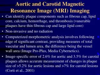
Magnetic Resonance Angiography The electromagnetic energy that is released when exposing a patient to radiofrequency waves in a strong magnetic field is measured and analyzed by a computer Most often used for evaluating the extracranial carotid arteries. Utilize either three dimensional time-of-flight MRA or gadolinium-enhanced MRA (contrast enhanced MRA).
Advantages Great imaging w/o use of contrast or radiation CEMRA – higher quality image with less artifact No catheter in area of interest May be most accurate non-invasive method Disadvantages May overestimate degree & length of stenosis? Use NASCET CI for pt’s with metallic implants CI in patients with renal insufficiency Magnetic Resonance Angiography
Cerebral Angiography • Cerebral angiography is the gold standard for imaging the carotid arteries. • The development of intraarterial Digital Subtraction Angiography (DSA) has largely replaced conventional angiograpy • Lower dose of contrast • Small catheters • Shorter procedure
Advantages Evaluates entire carotid a. system Information about the disease process Assess collaterals Disadvantages Invasive Expensive Radiation exposure Potential for stroke Limited views of carotid & bifurcation Cerebral Angiography
<50 % > 50% CEA more beneficial for asymp men with 60-99% stenosis who are good surgical candidates
May be benefit with 50 to 69% symptomatic stenosis (clearly shown in men but not women)
References • Up to date Online. Pathophysiology of Symptoms from Carotid Artery Atherosclerosis. Last revised May 1, 2006. • Up to Date Online. Evaluation of Carotid Artery Stenosis. Last revised June 22, 2006. • Rothwell, PM, Gibson, RJ, Slattery, J, et al. Equivalence of measurements of carotid stenosis. A comparison of three methods on 1001 angiograms. Stroke 1994; 25:2435. • Zwiebel, WJ. Duplex sonography of the cerebral arteries: Efficacy, limitations, and indications. AJR Am J Roentgenol 1992; 158:29. • Bowen, BC, Quencer, RM, Margosian, P, Pattany, PM. MR angiography of occlusive disease of the arteries in the head and neck: Current concepts. AJR Am J Roentgenol 1994; 162:9.