Download
1 / 31
320 likes | 974 Views
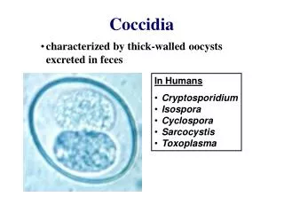
Coccidia of Small Animals. I. canis 35-45u I. ohioensis 20-30u I burrowsi 17-22u I. neorivolta 20-30u I. bigemina * 10-12u Neospora caninum 10-12u Sarcocystis spp 10-12u Cryptosporidium 4-6u

E N D
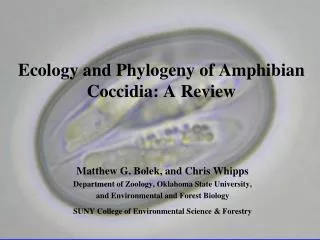
Coccidia of Small Animals
I. canis 35-45u I. ohioensis 20-30u I burrowsi 17-22u I. neorivolta 20-30u I. bigemina* 10-12u Neospora caninum 10-12u Sarcocystis spp 10-12u Cryptosporidium 4-6u *(=Hammondia heydorni) I. felis 35-45u I. rivolta 20-30u Hammondia hammondi* 10-12u Besnoitia* 10-12u Sarcocystis* 10-12u Toxoplasma 10-12u Cryptosporidium 4-6u * Assume Toxoplasma unless know otherwise Isospora (Cystisospora), other coccidiaDog Cat
Comparison of (clockwise) of Isospora felis, I. rivolta, Toxoplasma and Sarcocystis of cats; size ranges similar for dog coccidia
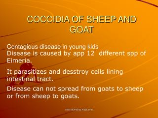
All Isospora of dogs and cats: • 2 sporocysts with 4 sporozoites • Intestinal cycle with monozoic cysts in tissues (paratenic hosts have only monozoic cysts • ~ One week prepatent period • High host specificity
Banana-shaped sporozoite from excysted sporulated oocyst. These invade intestinal epithelial cells to initiate schizogony or invade other tissues (eg. Lymph nodes) as monozoic cysts
Developing schizont (Meront)in intestinal epithelium cell • Schizogony=Merogony, asexual reproduction • 1-4 schizogony cycles merozoites
Gametogony (sexual reproduction) • Macrogametocyte fertilized by microgamete zygote Oocyst
Most exposed early in life Immune Massive exposure bloody diarrhea, enteritis Especially ‘puppy mills’, pet shops Main factors: number of oocysts exposure and immune state Often follows immunosuppression (eg shipping stress, distemper) Sulfa drugs (eg Albon) Furozolidone Amprolium prophylaxis or treatment prior/after weaning/shipping Supportive Rx until immunity builds Prepatent period – one week Self-limiting; maximum oocysts shed for 1 week, greatly reduce or cease in 2 week patent period unless immunosuppressed Clinical signs Treatment
Sporulated oocysts of Toxoplasma gondii • Oocysts shed by cats only (and other felidae) • Shed 10 million oocysts beginning 5-14 days after infection • Self-limiting after patent period of 2-3 weeks • Cats do not shed on second exposure, thus ‘safe’ (unless immunosuppressed) • Sporulate to infective form in 2 days -- change cat litter 1x/day; gardeners beware; oocysts survive 1 year in moist, cool; Cat is pivotal factor as source of herbivore infections
Sources of Toxoplasma infection in Cats: • Carnivorism (via bradyzoites, tachyzoites) – most efficient route; high % of cats shed large number of oocysts • Oocysts - 50% of cats shed small number of oocysts • Congenital – Clinical signs seen in offspring, especially if 1st trimester exposure • Cats develop both 1) the enteroepithelial phase of schizogony, gametogony in gut with oocyst shedding, and 2) the extraintestinal tissue phase of other hosts. Toxoplasma is an obligate intracellular, pantropic organism that can infect any warm-blooded vertebrate (including birds). In the extraintestinal phase, sporozoites, bradyzoites or tachyzoites invade lymph nodes, disseminate via lymph or blood to liver, lung then other tissues.
Rapid, proliferative schizogony (tachyzoites) occurs +/- clinical disease in intracellular ‘pseudocysts’ until contained by immunity in 7-10 days. The organism then persists long-term asymptomatically as ‘bradyzoites’ especially in reticuloendothelial system cells of the liver, lung, brain, eye, lymph nodes. When only partially contained, periodic recrudescence of multiplication, necrosis of tissues occurs, producing ‘chronic’ disease with signs depending on tissue location of organisms and lesions. • Congenital infections occur by invasive, proliferative tachyzoites in the potentially acute phase of the disease.
Toxoplasma is found in highest prevalence in moist environments in the USA and globally.
The cat is the key epidemiological factor in infection of herbivores. Surveys reveal about 1% of pound cats are shedding Toxoplasma oocysts at any one time. Sheep may abort or produce weak lambs if exposed during pregancy, but only once, then are immune. Placentitis
Carnivorism, not oocyst exposure, is the most important source of infection of humans: Beef< chicken < horse < lamb/goat < pork • Cattle are relatively resistant hosts and organisms are difficult to isolate from tissues, does not persist more than a few months; Filet mignon is OK? • Chicken is usually frozen or well cooked; Horsemeat not eaten in US • Lamb/goats – 5% of market lambs infected; can pass in goat milk • Pork – 1/3 infected in US surveys; 6% of hamburger contaminated by added pork, butcher equipment; Rare burgers have risk
Prevention in Food Sources: • Heat to over 70 C • Experimental irradiation, specialty market - 30Krad for pork kills organism and many bacteria • Meal preparation: Wash hands after handle raw meat; one cyst 100,000 bradyzoites • Salting, curing kills • Freezing with home freezer temperatures (-40F) kills but not reliable
Public Health: • Surveys reveal 1/3 people have titers to Toxoplasma, increases with age; Usually immune to new infection; May recrudesce with fatal results if immunosuppression (eg AIDS patients) • Humans usually develop asymptomatically or have flu-like symptoms in the acute phase; Death possible but most recover completely; some develop low-grade seething, recurrent chronic disease, especially eye, brain, liver, lung, lymphatic tissues. Location determines signs. Toxoplasma is thus often on differential diagnosis lists • If infection of non-immune mother occurs in pregnancy, 20-50% of fetus will be infected, some will abort • Most damage to fetus if infected in the first trimester Anencephaly, blindness, retardation, etc. Toxoplasma estimated to occur in 1 of 1000 births, most are asymptomatic, some delay signs for years (eg. until puberty) • Number of infective stages ingested, immune state are the crucial factors in infection course
Diagnosis • Oocysts in cats can be found by flotation procedures at 5-24 days post infection only • Sheathers sugar (5 lb sucrose + 1450 water + 29 g phenol) is best • Do ‘Double Flotation’ – float 10 minutes, coverslip, let float to under coverslip 10 minutes; film is 100u, Toxoplasma only 10-12u; Focus just under coverslip
Serology: • Reliable human, dog, other species • Cats notorious for developing low, unreliable titers after 4-6 weeks • Sabin-Feldman Dye test (oldest test) • IFA: Common use; IgM early • ELISA: Detect IgM, IgG, Antigen • IHA, CF: Late IgG, not as sensitive • Do paired sera 2-3 weeks apart – 4x rise in titer is diagnostic; >1:256 considered positive for active infection, especially cats; 1:1024 is the cut-off by many for clinical disease • There are many different strains for Toxoplasma (and other protozoan diseases)
Animal inoculation • Animal inoculation can be done using oocysts or tissue (pepsin digest + wash) with oral, intraperitoneal or intracranial infection of mice, rats. • Find organism in peritoneal fluid in 4-5 days, in tissues 4-6 weeks, by serology in 3 weeks Peritoneal fluid Impression smear Brain section
Treatment • Sulfa drugs: Sulfadiazine (60mg.kg.day) + Pyrimethamine (0.5-1.0 mg/kg/day) + yeast or folinic acid (for side effects), or long acting sulfa drugs (eg Tribrissen, Di-Trem) • Clindamycin is drug of choice
Classic predator-prey life cycle, numerous species • High host specificity • Gametogony + sporogony in predator, Schizogony tissue phase in prey • Acute disease possible in proliferative schizogony phase (counterpart of tachyzoites) in reticuloendothelial tissues • Skeletal and cardiac muscle tissue stage is quiescent (counterpart of bradysoites) • May shed high numbers for extended periods (eg 60 days); little gut pathogenicity; No immunity to re-infection develops in predators • Human ‘Isospora’ now known to be S. bovihominis or S. porcihominis
Hammondia • Indistinquishable from Toxoplasma (10-12u) • Non-pathogenic in cats which have intestinal gametogony phase only; Schizongony in muscle tissue of rodent hosts; Sporogony in the environment
Besnoitia • Slightly larger than Toxoplasma (12-17u) • Asymptomatic gametogony in cat, sporogony in environment, schizogony in subcutaneous tissues of rodent, herbivores, etc • Differential diagnosis importance only
Cryptosporidium • Occurs in dogs and cats, but low prevalence • Probably of minor clinical or public health importance • Differentiate from yeast, which are not acid fast and are variable in size
Neospora caninum • Newly described in dog (1988);previously confused with Toxoplasma • Oocysts 10-12u in size are shed by dogs; Source of infection for abortion in dairies, beef cattle • Ultrastucture, immunohistochemistry different from Toxoplasma • Clinical disease may result in dogs which is similar to Toxoplasmosis, but neurological and muscular abnormalities predominate with Neospora
Pneumocystis carinii • Organism of uncertain taxonomy (yeast vs sporozoan) that infects respiratory tissues of humans, dogs, other species, most subclinical , <10% dry non-productive cough • If immunosuppress, proliferate in alveoli with clinical disease • Diagnosis by transtracheal wash, biopsy or sputum sample ID organism; By histopathology, does not stain with Hemotoxylin and Eosin; must use special stains including Giemsa, Wrights, Gram stain or Silver stain (the latter is best) • Treatment may be tried with sulfas (eg Tribrissen), pentamidine isothionoate