Download
1 / 44
2.69k likes | 12.45k Views
Dental pulp. Dr. Fatma Elturki. Objectives. Definition of the dental pulp. Development of the pulp. Anatomy of the pulp. Histology of the pulp. Functions of dental pulp. Age changes of dental pulp. Clinical considerations. Dental pulp. It is loose delicate connective tissue.

E N D
Dental pulp Dr. Fatma Elturki
Objectives • Definition of the dental pulp. • Development of the pulp. • Anatomy of the pulp. • Histology of the pulp. • Functions of dental pulp. • Age changes of dental pulp. • Clinical considerations.
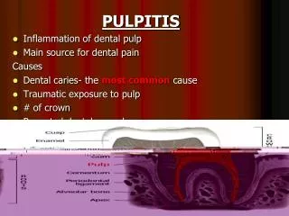
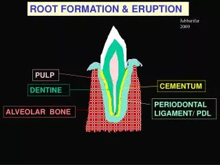
Dental pulp • It is loose delicate connective tissue. • It occupying the cavity lying in the center of dentin. • It is confined within the pulp chamber and root canals of the tooth.
Pulp development • The pulp derived from ectomesenchymal cells of dental papilla. • During dentinogenesis, when the dentin forms around the dental papilla, the innermost tissue is considered pulp.
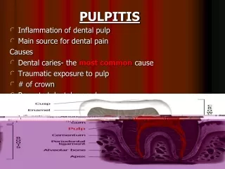
Anatomy of the pulp Apical foramen: • The root canal terminate at the apical foramen, where the pulp and PDL meet and the main nerves and vessels enter and leave the tooth. • In the developing tooth the apical foramen is large and centrally located. • As the tooth completes its development, the apical foramen become smaller in diameter and more eccentric in position.
Anatomy of the pulp Accessory or lateral canals: • Connections between the pulp and PDL may also occur along the lateral surface of the root through accessory canals.
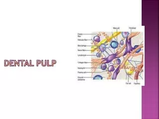
Histology of the pulp • The odontoblastic Zone: On the periphery of the pulp, adjacent to the calcified dentin, Next to the predentin lies the palisade of columnar odontoblast cells. • The cell-free zone (zone of Weil): beneath the odontoblasts in the coronal pulp. • cell-rich zone: where cell density is high. • Pulp core: The main body of the pulp occupies the area circumscribed by cell-rich zones. It contains the principal support system for the peripheral pulp, which includes the large vessels and nerves.
Structural Elements • Cells: which includes: • Synthetic cells (fibroblasts, odontoblasts). • Defensive cells (Dendritic cells , macrophages, T-lymphocytes, etc). • Progenitor cells (undifferentiated mesenchymal cells). • Fibers. • Ground substances. • Blood supply. • Lymphatics. • Nerve supply.
Synthetic cells 1- Fibroblasts • Fibroblasts are the most abundant cell population of the pulp tissue. • They synthesize and secrete the bulk of the extracellular components, that is collagen and ground substance. • In addition to synthesis of collagen, they also eliminate excess collagen or participate in collagen turnover in the pulp by resorption of collagen fibers. • In young pulp, the fibroblasts appear large with multiple processes, centrally placed nucleus, with all protein synthesis organelles. By age, the cell become smaller spindle shaped with few organelles.
Synthetic cells 2-Odontoblast • Odontoblasts are the 2nd largest group of cells in the pulp. • It is the principal cell of the dentin forming layer • It is located around the pulp chamber as a single cell layer. But only their cell bodies are located lining the outer pulpal surface and their process extend in the DT in dentin.
Defensive cells Histocytes and Macrophages:- • These cells are highly phagocytic and can remove bacteria, foreign bodies (endodontic paste, zinc oxide, etc), dead cells, or other debris.
Defensive cells Polymorphonuclear Leukocytes:- • The most common form of leukocyte in pulpal inflammation is the neutrophil, although eosinophils and basophils are occasionally detected. • It is important to know that although neutrophils are not normally present in intact healthy pulps, with injury and cell death they rapidly migrate into the areas from nearby capillaries and venules.
Defensive cells Pericytes or endothelial cells:- • They located around blood vessels. The function of these cells is controversial, but they are though to be contractile cell capable of reducing the size of the vessel lumen. • It may differentiate into odontoblasts.
Defensive cells Lymphocytes and Plasma Cells:- • These inflammatory cell types generally appear following invasion into the area of injury by neutrophils. • These cells are not normally present in healthy pulp tissue but are associated with injury. • Their presence would therefore indicate the presence of a persistent irritant.
Defensive cells Mast Cells:- • These cells are seldom in large numbers in normal, healthy pulps but are commonly found in inflamed pulps. • The granules of these cells contain histamine, and heparin. • These cells release these granules into the surrounding tissue fluid during inflammation and they are generally found near blood vessels.
Defensive cells Dendritic cells:- • Bone marrow-derived, antigen presenting dendritic cells. • They stimulate the division and activity of T-lymphocyte. • Found in and around the odontoblast cell layer.
Progenitorcells Undifferentiated Ectomesenchymal cells:- • It is embryonic branched cell & can be differentiated into other types of connective tissue cells. • It is smaller than fibroblasts but have similar appearance. • They are usually found along the walls through out the cell rich zone and pulp core. • With age they decrease in number.
Structural Elements Extracellular:- • The dental pulp has most of its volume primarily composed offibers and ground substance. • These form the body and integrity of the pulp organ.
Extracellular Fibers:- • These fibers form a loose, reticular network to support other structural elements of the pulp. • The fibers are principally type I and type III collagen .
Extracellular ground substance :- • This structureless mass, gel-like in consistency, makes up the bulk of the pulp organ. • It resembles the ground substance of any connective tissue . • Composed mainly by combinations of Glycosaminoglycans, glycoproteins and water.
Supportive Elements Pulpal Blood Supply: - • The pulp is highly vascularized, with vessels arising from the external carotids to superior and inferior alveolar arteries and drains by the same veins. • Capillary density is highest in the subodontoblastic region with loops passing between odontoblasts In the subodontoblastic region, • capillaries with fenestrations occur frequently and regularly in both primary and permanent teeth.
Supportive Elements Lymphatics:- • Lymphatic vessels have been identified in the pulp at the ultra structural and histologic levels by the absence of red blood cells in their Lumina, and the absence of a basal lamina.
Supportive Elements Nerves:- • Several nerve bundles, each containing numerous unmyelinated and myelinated nerves, pass into each root via the apical foramen. • The myelinated nerve fibers branch extensively beneath the cell-rich zone to form the so-called plexus of Raschkow. • many fibers lose their myelin sheath and pass through the cell-free zone to terminate as receptors or as free nerve endings near odontoblasts.
Functions of dental pulp 1. Formative. formation of dentin by odontoblast cells during the developmental period. 2. Nutritive. the high blood supply of the dental pulp transfer the nutrients to the tooth. 3. Sensory. the complex sensory system within the dental pulp controls the blood flow and is responsible for at least mediation of the sensation of pain. 4. Defensive. formation of reparative or secondary dentin represents a defensive response to any form of irritation.
Age changes of dental pulp 1- The most conspicuous changes is decreasing volume of the pulp chamber and root canal due to continuous dentin deposition.
Age changes of dental pulp 2- With increasing age the pulp tissue shows a reduction in the number of the cellular elements specially progenitor cells, leading to reduction in regenerative potential of the pulp. Young pulp Old pulp
Age changes of dental pulp 3- The collagen fibers increase in number and density which becomes more evident with the decrease in pulp size, so it produce an actual fibrosis.
Age changes of dental pulp 4- with age, there is both loss and degeneration of myelinated and unmyelinated nerve fibers that correlated with age- related reduction in sensitivity.
Photomicrograph of control group after six months showing nerves plexus under the odontoblastic zone and nerve bundle in the center of the pulp core (Silver stain, origin. Mag. ×100)
Photomicrograph of control group after 12 months showing nerve bundles in root canals (silver stain, origin. Mag.×400).
Age changes of dental pulp 5- Calcification (pulp stones) is a common age changes of the pulp that may be occurs as localized or diffuse pulp calcification. Pulp stones or denticles are nodular, calcified masses appearing in either both the coronal and root portions of the pulp. They are usually symptomless unless they impinge on nerves or blood vessels.
Diffuse calcifications • Diffuse calcification appear as irregular calcific deposits in the pulp tissue, usually following collagenous fiber bundles or blood vessels. • Diffuse calcifications are usually found in the root canal and less often in the coronal area.
Clinical considerations 1- With advancing age, there is a difficulty in the endodontic treatment due to: • Excessive dentin formation at the roof and floor of the pulp chamber. • Presence of pulp stones at the opening of the root canal. • The apical foramen is narrowed by cementum.
Clinical considerations 2- when accessory canals are located near the coronal part of the root or in the bifurcation area, a deep periodontal pocket may cause pulp inflammation. Conversely a necrotic pulp can cause periodontal disease.
Clinical considerations 3- A non vital tooth become brittle and is subjected to fracture. Therefore, every precaution should be taken to preserve the vitality of the pulp.
Clinical considerations 4- some materials such as calcium hydroxide seem to facilitate dentin bridge formation. So, they applied in deep cavity preparation when the dentin layer is very thin.