
Introduction
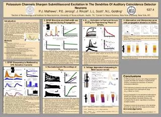
A. -20mV. -90mV. -60mV. V pk -V rest. V pk -V rest. V pk -V rest. V pk -V rest. Potassium Channels Sharpen Submillisecond Excitation In The Dendrites Of Auditory Coincidence Detector Neurons P.J. Mathews 1 , P.E. Jercog 2 , J. Rinzel 2 , L.L. Scott 1 , N.L. Golding 1

Introduction
E N D
Presentation Transcript
A -20mV -90mV -60mV Vpk-Vrest Vpk-Vrest Vpk-Vrest Vpk-Vrest Potassium Channels Sharpen Submillisecond Excitation In The Dendrites Of Auditory Coincidence Detector Neurons P.J. Mathews1, P.E. Jercog2, J. Rinzel2, L.L. Scott1, N.L. Golding1 1Section of Neurobiology and Institute for Neuroscience, University of Texas at Austin, Austin, TX, 2Center for Neural Science, New York University, New York, NY. 637.4 2. EPSP Rise-time and Half-width are Maintained During Propagation 4. K(LVA) Activation is Fast and Occurs During the Repolarizing Phase of EPSPs 5. Attenuation and Sharpening grow with propagation distance in model. Introduction In mammals, binaural auditory signals are integrated in the dendrites of principal neurons of the medial superior olive (MSO). Since the temporal resolution of this integration is directly related to the acuity of azimuthal sound localization, a critical issue is how synaptic precision is maintained in the face of temporal distortions introduced by dendritic cable properties. To address this question, we combined somatic and dendritic patch recordings from MSO principal neurons (brainstem slices, 16-20 day gerbils) with compartmental modeling.Our findings show that (K(LVA)) helps to maintain the brevity and fidelity of dendritically propagating EPSPs by preventing stereotypical broadening associated with dendritic filtering. b a a b The Binaural Circuitry Coronal view of the binaural circuitry in the ventral brainstem. The MSO integrates binaural excitatory and ipsilateral inhibitory synaptic inputs. VCN, ventral cochlear nucleus; MNTB and LNTB, medial and lateral nucleus of the trapezoid body, respectively. EPSC amplitude fixed. Input at => EPSP of 12mV at soma. • Reduction of gKLVA (“DTX”) converts sharpening to broadening and reduces attenuation. • Distance dependence of sharpening matches experimental observations if V-activation of gKLVA is steeper in dendrites. c c d • Methods • General • Mongolian gerbils, 14-23 days old • Horizontal brainstem slices, 200 µm thick, maintained at35° C during recordings • Neurons visualized using infrared DIC microscopy Bilateral stimulation enhances sharpening B C • Current Clamp Recordings • Whole-cell somatic and dendritic recording configurations were carried out using capacitance neutralization and bridge balance compensation • Somatic patch pipettes were 3-4 MΩ (somatic) and 6-10MΩ (dendritic) • Synaptic stimulation with glass patch pipettes with tips broken to ~20-50 µm • Kgluconate-based internal solution with 0.5 mM EGTA • Nucleated-Patch Voltage Clamp Recordings • Kgluconate-based internal solution with 5.0 mM EGTA • Na+, high threshold K+, Ca+ and h channels were blocked with 1 µM TTX, 10 mM TEA-Cl, 200µM Co2+ and 50 µM ZD7288 (bath applied). AMPA receptors blocked with 10 µM CNQX • Patch electrodes: 2-3 MΩ resistances and access resistances of < 8 MΩ • Electrodes were wrapped with parafilm to decrease pipette capacitance Stimulus Peak • Somatic EPSPs from different dual dendritic (orange) and somatic (blue) recordings elicited with simEPSCs adjusted to be just below action potential threshold. Amplitude of current injections: 2.3, 3.2, and 2.8 nA for dendritic recordings 35, 55, and 73 µm from the soma respectively. • Group data from all paired recordings, showing dendritic responses (orange circles) and somatic responses (blue circles). Closed circles at 0 µm: somEPSP responses to somatic current injection in dual somatic recordings. • C. Absolute value of somEPSP rise time and halfwidth as a function of the location of current injection along the somatodendritic axis. Linear fits both exhibit slopes of –0.001 ms/µm and are constrained to pass through the average somatic responses at 0 µm (rise time: 0.39 ms, halfwidth: 0.77 ms). Activation of TEA-resistant potassium conductances during voltage steps (-90 mV holding potential to voltages between -60 and –20 mV, in 10 mV increments). Plot of activation time constant (tauact) vs. voltage step. Activation of outward potassium currents with EPSP waveforms. Patches were held at –90 mV to remove inactivation, and subjected to an EPSP waveform that was scaled to reach different peak voltages. Summary data showing the timing of potassium current peak (blue) and foot (orange) relative to the peak of EPSP commands (t). The foot was defined as the point at which the beginning of the current was 2 standard deviations above the noise. Passive model with gKLVA frozen at rest (far right panel) shows slow decaying EPSPs (note different time scale). 1. EPSP Sharpening is Mediated by DTX Sensitive Currents 3. Nucleated-patch Recordings of K(LVA) 4. Voltage dependent sharpening in MSO neuron model. b a a b Neuron Model • Weak INa, ( set Ina = 0 ). • Kinetics set for 35˚C. • Typically, m = 0.6ms, Rin=7-8M as in experiments. • differs in S and D: in nucleated patch, = 4.5pS/µm2 • EPSC as alpha fn., = 0.2ms. • uniform in D • Each dendrite (150µm), electrotonic length of 1 , modeled with 10 compartments • Conclusions • In MSO principal neurons, low voltage-activated K+ currents decrease the duration of subthreshold EPSPs, increasing the resolution of binaural coincidence detection. • TEA insensitive potassium channels located at the soma activate in the subthreshold range (V1/2=-35mV, k=17mV). • During propagation in the dendrites subthreshold EPSPs are actively shortened to counteract the broadening effects of dendritic filtering, and normalize their duration regardless of excitatory synaptic location. • Potassium channels are present in the dendrites and can be resolved at the single channel level. • Acknowledgements • This work was supported from NIH grant DC006341 ??? IKLVA IH ILEAK S, soma c d D, dendrite c f e EPSC Simultaneous dendritic (orange traces) and somatic (blue traces) voltage responses to a family of simulated EPSCs (simEPSC) recorded in control ACSF and in the presence of 100 nM -DTX. simEPSCs were injected in the lateral dendrite 90 µm from the soma (schematic inset). Quantification of the effects of DTX on EPSP halfwidth. Normalized traces of 3 selected somatic EPSPs (somEPSP) in A, showing the voltage-dependent sharpening over the full subthreshold voltage range (left). EPSP sharpening is abolished in the presence of 100nM -DTX (right). Group data showing the degree of voltage-dependent sharpening at the soma when simEPSCs were injected in the soma (“Local”, left graph, n=5), or in the dendrite 55-90 µm away (“Propagated”, right graph, n=4). Normalized somatic (blue traces) and dendritic (orange traces) EPSPs produced by simEPSCs in the dendrite with and without 100nM -DTX. Group data showing the increase in duration in the presence of 100 nM -DTX (n=x). Superimposed outward potassium currents (top traces) under voltage clamp conditions to a series of steps from a holding potential of –90 mV to tests potentials between –70 mV to +40 mV (10 mV increments). The remaining TEA-insensitive outward currents were extensively blocked in the presence of 100 nM -DTX (middle traces). The DTX-sensitive current was obtained via the subtraction of currents recorded in DTX from control traces (bottom traces). High voltage-activated potassium, Na+ , Ih and Ca2+ currents blocked with TEA (10mM), TTX (1µM), ZD7288 (50µM) and CoCl (200µM), respectively. Current-voltage relationship for the potassium currents in A. Plot of normalized conductance versus voltage (V1/2=-35mV, k=17mV, n=3). Plot fitted to the Boltzmann equation, f(V)=1/(1+exp((V1/2-V)/k)). Plot of normalized conductance vs. voltage for all patches where reliable DTX subtractions were performed (n=5). Halfwidth • Left: Weak in S and D => broadening not sharpening. • Center: Stronger in D => modest sharpening. • Right: Steeper activation (Rothman and Manis, 2003) in D => substantial sharpening.


