
Introduction
The plasma needle: A non-thermal atmospheric plasma for treatment of biological tissue Ingrid Kieft , Raymond Sladek, Eva Stoffels Department of Biomedical Engineering, Eindhoven University of Technology P.O.Box 513, 5600 MB Eindhoven, The Netherlands E-mail: I.E.Kieft@tue.nl. Introduction.

Introduction
E N D
Presentation Transcript
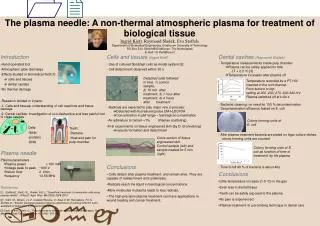
The plasma needle: A non-thermal atmospheric plasma for treatment of biological tissue Ingrid Kieft, Raymond Sladek, Eva Stoffels Department of Biomedical Engineering, Eindhoven University of Technology P.O.Box 513, 5600 MB Eindhoven, The Netherlands E-mail: I.E.Kieft@tue.nl Introduction Cells and tissues (Ingrid Kieft) Dental cavities (Raymond Sladek) • Temperature measurements inside pulp chamber • Plasma can be safely applied for 60s: • T < 2.2 oC [3] • Temperature increases after plasma off • Hand-operated tool • Atmospheric glow discharge • Effects studied in biomedical field [1] • cells and tissues • dental cavities • No thermal damage • Use of cultured fibroblast cells as model system [2] • Cell detachment observed within 30 s Detached cells followed in time. 1) control sample, 2) 15 min. after treatment, 3) 1 hour after treatment, 4) 4 hours after treatment 1 2 Temperature recorded by a PT-100 inserted in the root channel. From bottom to top: setting at 200, 230, 270, 300,340 mV Plasma switched off at t=60 s 3 4 • Research divided in 2 parts: • 1. Cells and tissues: understanding of cell reactions and tissue damage • 2. Dental cavities: Investigation of non-destructive and less painful tool to clean cavities • Bacterial cleaning: no need for 100 % decontamination • Decontamination efficiency tested on E. coli • Radicals are expected to play major role in process: • Detected with fluorescent probe CM-H2DCFDA • Concentration in µM range ~ fysiological concentration • Air admixture to helium <1% (Raman scattering) • First experiments on tissue engineered skin (by D. Bronneberg) • vacuole formation and detachment Colony forming units of E. coli • Cells: • lipids • proteins • DNA • Teeth: • Bacteria • Heat and pain for pulp chamber • After plasma treatment bacteria are plated on Agar culture dishes, • colony forming units are counted Cross section of tissue engineered skin. Control sample (left) and sample treated for 5 min (right) Colony forming units of E. coli as function of time of treatment by He plasma Plasma needle • Plasma parameters • Plasma power < 100 mW • Voltage peak to peak ~600 V • Helium flow 2 l/min • frequency 13.56 MHz • Conclusions • Cells detach after plasma treatment, and remain alive. They are capable of reattachment and cytokinesis. • Radicals reach the liquid in fysiological concentrations. • More molecules in plasma leads to less radicals. • The high precision plasma treatment can have applications in wound healing and cancer treatment. - Time to kill 90 % of bacteria is about 40s. • Conclusions • Little temperature increase (1-5 oC) in the gas • Even less in dental tissue • Teeth can be safely exposed to the plasma • No pain is experienced • Plasma treatment is a promising technique in dental care References: [1] Stoffels,E., Kieft, I.E., Sladek, R.E.J., “Superficial treatment of mammalian cells using plasma needle”, J.Phys.D: Appl. Phys. 36 (2003) 2908-2913 [2] Kieft, I.E., Broers, J.L.V., Caubet-Hilloutou, V., Slaaf, D.W., Ramaekers, F.C.S., Stoffels, E., “Electric discharge plasmas influence attachment of cultured CHO K1 cells”, accepted for publication in Bioelectromagnetics [3]Zach, L. and Cohen, G., “Pulp response to externally applied heat”, Oral Surgery, Oral Medicine, Oral Pathology, 19, no. 4, (1965) 515-530


