Download
1 / 1
10 likes | 97 Views
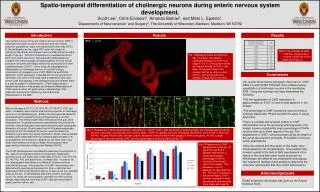
The study investigates the spatio-temporal localization of cytoskeletal filaments during auditory organ (organ of Corti) development in rats from embryonic day 18 to post-natal day 25. Results reveal distinct patterns of actin and tubulin isoforms present in sensory and supporting cells, with intermediate filaments mainly in supporting cells. The study sheds light on how cytoskeletal proteins vary across cellular types and developmental stages, with significant changes seen between post-natal days 8 and 12.

E N D
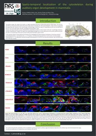
Spatio-temporal localization of the cytoskeleton during auditory organ development in mammalia Nicolas JOHNEN, Marie Cloes, Nicolas Thelen and Marc Thiry GIGA-Neuroscience, Cell and Tissue biology Unit, Université de Liège Introduction The auditory organ, the organ of Corti (OC), is a highly specialized structure composed by specific cellular types. The sensory cells (HC) are characterized by stereocilia at their apex and are necessary for the sound perception. These cells are supported by supporting cells. Based on their morphology and physiology, at least four types of supporting cells (SC) can be identified in the OC: inner and outer pillar cells (PC), phalangeal cell and Deiter’s cells. Sensory and supporting cells possess characteristic cytoskeleton proteins in direct relation with their morphological features and their development. Indeed, this organ had morphological changes such as the setting up of the sensory epithelium after the birth or the openings of the Corti’s tunnel at P8 and of the Nuel’s spaces at P10. In the present study, by using confocal microscopy, we investigated the spatio-temporal localization of the three cellular cytoskeletal filaments : microtubules (β-1, 2, 3, 4-tubulin), microfilaments (cytoplasmic β- and γ-actin) and intermediate filaments (CK4, 5, 7, 8, CKpan and vimentin) during the development of the OC in rat from the embryonic day 18 (E18) to the post-natal day 25 (P25). Results Actinβ Actinγ βTubulin I βTubulin II βTubulin III β Tubulin IV Cytokeratine 8 Vimentin Figures AB, G1-AB, G6/ BTI, II, III, IV1- BTI, II, III, IV6/ CK8 1-CK8 6/ Vim 1-Vim 6: Spatiotemporal distribution of cytoplasmic β-actin isoform (AB)/ cytoplasmic γ-actin isoform (AG)/ Beta-Tubulin I, II, III and IV (BT)/ cytokeratin 8 (CK8)/ Vimentin (Vim) during the mammalian auditory organ development from E21 to P16 in the basal part of the cochlea, respectively. The immunolabellings indicated clearly that β-1, 2, 3-tubulins were only present in the SC and nervous fibers during development whereas β-4-tubulin was found firstly in the HC and then in the SC. The two actin-isotypes were detected in the HC apex but were also seen in the PC from P8 to P25 for β-actin isoform and in the basal membrane from E18 to P8 for the γ-actin isoform. All intermediate filament proteins were only found in the SC, especially between P8 and P12. IH: Inner hair cell; OH: Outer hair cell; IP: Inner Pillar cell; OP: Outer Pillar cell; D: Deiters' cell; Ip: Phalangeal cell. Bar: 10µm. Red: Cytokeratin 8 or Myosin VI; Green: Cytoplasmic β/ γ-actin isoform or Beta-tubulin I-IV or Vimentin/p27kip1; Blue: Dapi. Conclusion Our results show that the localization of the cytoskeleton proteins during the auditory organ development depends on the cellular type and the developmental stage. A profound modification of cytoskeleton occurs between P8 and P12 Contact : n.johnen@ulg.ac.be