Download
1 / 33
330 likes | 456 Views
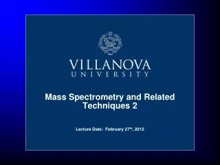
Mass Spectrometry and Related Techniques 2. Lecture Date: February 27 th , 2012. Mass Analyzers - Outline. Sector Mass Analyzers (Magnetic and Electrostatic) Quadrupole Analyzers Ion Traps Ion Cyclotron Resonance Time-of-Flight and many more…. Ionization Source. Mass Analyzer.

E N D
Mass Spectrometry and Related Techniques 2 Lecture Date: February 27th, 2012
Mass Analyzers - Outline • Sector Mass Analyzers (Magnetic and Electrostatic) • Quadrupole Analyzers • Ion Traps • Ion Cyclotron Resonance • Time-of-Flight • and many more…. Ionization Source Mass Analyzer Detector
Properties of Mass Analyzers Resolution (R): R = m/m m = mass difference of two adjacent resolved peaks m = mass of first peak or average Example: R = 500 (“low” resolution) resolves m/z=50 and 50.1, and m/z=500 and 501 Example: R = 150000 (“high” resolution) resolves m/z=50 and 50.0003, and m/z=500 and 500.0033
Sector Mass Analyzers • Basic Features • A sector is a geometrical construction that consists of two arcs inside of one another. • Types: • Magnetic • Electrostatic • Combination (e.g. double-focusing)
Magnetic Sector Mass Analyzers Ion kinetic energy: Where: T is kinetic energy z is charge on ion e is electron charge (1.60 x 10⁻¹⁹ C) B is magnetic field (T) v is velocity (m/s) V is the accelerating voltage m is the mass Forces: Only ions with equal forces will pass: Therefore: Diagram from Strobel and Heineman, Chemical Instrumentation, A Systematic Approach, Wiley, 1989.
Electrostatic Sector Mass Analyzers Ion kinetic energy: Forces: Only ions with equal forces will pass: V can be varied to bring ions of different KE (and different m/z ratio to the exit) Therefore: Diagram from Strobel and Heineman, Chemical Instrumentation, A Systematic Approach, Wiley, 1989.
Double-Focusing Sector Mass Analyzers • If a batch of ions of equal m/z but with different kinetic energies enters a magnetic sector instrument, this will result in a spread-out beam • Soution: minimize directional and energy differences between ions of the same m/z. • Example of a double-focusing MS: the Nier-Johnson geometry Diagram from Strobel and Heineman, Chemical Instrumentation, A Systematic Approach, Wiley, 1989.
Double-Focusing Sector Mass Analyzers • Another design, the Mattauch-Herzog geometry • This geometry is analogous to CCD-based optical electronic spectroscopy systems, while Nier-Johnson instruments are similar in nature to traditional scanning monochromator spectrometers. Diagram from Strobel and Heineman, Chemical Instrumentation, A Systematic Approach, Wiley, 1989.
Time-Of-Flight (TOF) Mass Analyzers • The principle of “Time-of-flight” mass analysis: • A batch of ions is introduced into a chamber by an pulse of accelerating current. • This chamber has no fields, and is a “drift tube” • Since the ions have the same kinetic energy, their velocities vary inversely with their mass during their drift. • Notes: • Typical flight times are 1-30 us • Lighter ions arrive at the detector first M. Guilhaus; Journal of Mass Spectrometry, 30; 1995, p1519.
Time-Of-Flight (TOF) Mass Analyzers • Delayed extraction – anything you can do to tighten the KE spread will help a TOF instrument m/z is mass-to-charge ratio of the ion E is the extraction pulse potential(V) s is the length of flight tube over which E is appliedd is the length of field free drift zonet is the measured time-of-flight of the ion M. Guilhaus; Journal of Mass Spectrometry, 30; 1995, p1519.
Time-Of-Flight (TOF) Mass Analyzers • The reflectron – a method of compensating for different ion KE’s Figure from http://www.abrf.org/ABRFNews/1997/June1997/jun97lennon.html
Time-Of-Flight (TOF) Mass Analyzers • The reflectron – a method of compensating for different ion KE’s Figure from http://www.abrf.org/ABRFNews/1997/June1997/jun97lennon.html
Quadrupole Mass Analyzers • The quadrupole (named for its “electrical structure”) is one of the simplest and most effective mass spectrometers. Diagrams from Skoog et al.
Quadrupole Mass Analyzers • How a quadrupole works: • Most important points: • It is easier for an applied AC field to deflect a light ion than a heavier ion • Conversely, it is easier for an AC field to stabilize a light ion • Using this knowledge – a combined AC/DC potential is applied to the rods. Via the DC, the ion is attracted to one set of rods and repelled by the other • The DC serves to stabilize heavy ions in one direction (high pass filter). The AC serves to stabilize light ions in the other direction (low pass filter). • The ion must pass through the quadrupole to make it to the detector Diagrams from Skoog et al.
Quadrupole Mass Analyzers • Another view – and the concept of the mass scan… Light ion: (ex. m/z = 100) Dragged by AC Heavy ion: (ex. m/z = 500) Dragged by DC Just right: Dragged by both, But equally balanced Images from http://www.jic.bbsrc.ac.uk/SERVICES/metabolomics/lcms/single1.htm
Ion Trap Mass Analyzers • Ion trap: a device for trapping ions and confining them for extended periods using EM fields • Used as mass analyzers because they can trap ions and eject them to a detector based on their mass. • Theory is based on Mattieu’s work on 2nd order linear differential equations (in the 1860’s), and on Wolfgang Paul’s Nobel Prize winning implementations R. E. March and R. J. Hughes, Quadrupole Storage Mass Spectrometers, Wiley, 1989. See also Chem. Eng. News 1991; 69(12):26-30, 33-41 Figure from W. Paul Nobel Lecture, December 8, 1989.
Ion Trap Mass Analyzers • The stability region of an ion trap – based on differential equations • Most ITMS systems don’t use DC (U), i.e. only qzis controlled R. E. March and R. J. Hughes, Quadrupole Storage Mass Spectrometers, Wiley, 1989.
Ion Trap Mass Analyzers • Layout of a cylindrical ion trap mass analyzer: Diagram courtesy of M. Olsen, GlaxoSmithKline
Ion Trap Mass Analyzers • The Thermo LTQ Velos Pro, a modern dual-pressure linear ion trap For more about dual-pressure linear traps, see J. V. Olsen et al., A dual pressure linear ion trap Orbitrap instrument with very high sequencing speed, Molecular and Cellular Proteomics, 8, 2759-2769. For more about linear traps, see D. J. Douglas et al., Linear ion traps in mass spectrometry, Mass Spectrometry Reviews, 2005, 24, 1–29
Ion Cyclotron Resonance • FT-ICR: a FT-based mass spectral method that offers higher S/N, better sensitivity and high resolution • Also contains a form of ion trap, but one in which “ion cyclotron resonance” occurs. • When an ion travels through a strong magnetic field, it starts circulating in a plane perpendicular to the field with an angular frequency c:
Ion Cyclotron Resonance • How ICR works: • The ions are circulated in a field • An RF field is applied to match the cyclotron frequency of the ions – this field brings them into phase coherence (forming ion “packets”)! • The image current is produced as these little packets of ions get near the plates. The frequency of the image current is characteristic of the ion packet’s m/z ratio. http://www-methods.ch.cam.ac.uk/meth/ms/theory/fticr.html
Ion Cyclotron Resonance and Magnetic Field • Parallels between NMR/EPR and ICR: B B ze B B = = m Picture courtesy Prof. Alan Marshall, FSU/NHMFL
The Orbitrap: A “Hybrid” Trap – Between IT and ICR • The Orbitrap is an electrostatic ion trap with FT/MS read-out of image current, coupled with MS/MS • Advantages • Ease of use • Resolving power (superior to TOF) • Precision and accuracy • Versatility, dynamic range • A lower-resolution, more economical ICR
LTQ Orbitrap schematic Finnigan LTQ™ Linear Ion Trap API Ion source Linear Ion Trap C-Trap Orbitrap Differential pumping Differential pumping Image/animation from Thermo Electron Inc. See A. Makarov et al., Anal. Chem.2006,78, 2113-2120.
LTQ Orbitrap Operation Principle 1. Ions are stored in the Linear Trap 2. …. are axially ejected 3. …. and trapped in the C-trap 4. …. they are squeezed into a small cloud and injected into the Orbitrap 5. …. where they are electrostatically trapped, while rotating around the central electrode and performing axial oscillation The oscillating ions induce an image current into the two outer halves of the orbitrap, which can be detected using a differential amplifier Ions of only one mass generate a sine wave signal Image/animation from Thermo Electron Inc. See A. Makarov et al., Anal. Chem.2006,78, 2113-2120.
Frequencies and Masses The axial oscillation frequency follows the formula Where w = oscillation frequency k = instrumental constant m/z = mass-to-charge ratio Ions in the Orbitrap generate a complex signal whose frequencies are determined using a Fourier Transformation Image/animation from Thermo Electron Inc. See A. Makarov et al., Anal. Chem.2006,78, 2113-2120.
Multiple-Stage MS: MS-MS, and MSn • Also known as Tandem MS or MSn … Mass Analyzer Mass Analyzer • Multiple quadrupoles are very common (e.g. triple-quad or QQQ systems, EB for double-focusing, Q-TOF for quad time-of-flight…) • Why tandem MS? Because of the possibility of doing CID – collisionally induced dissociation. Ions are allowed to collide with a background gas (He) for several millliseconds, prior to analysis. Allows for MSn experiments in an ion trap.
Comparison of Mass Analyzers • A comparison of the properties of some common mass analyzers
Detectors for Mass Spectrometry • Electron multipliers: like a photomultiplier tube. Ions strike a surface, cause electron emission. Each successive impact releases more electrons • Faraday Cups: Ions striking a cup cause charge to flow across a load. The potential across the load is monitored. • See pg 257 of Skoog et al. for more details. Ionization Source Mass Analyzer Detector Figure from D. W. Koppenaal, et al.; Anal. Chem., 77; 2005, 418A-427A.
Detectors: Electron Multipliers • Electron multiplier (EM): most common design in current use • High gain (107), low noise, good dynamic range (104-106) • Several designs: Figure from D. W. Koppenaal, et al.; Anal. Chem., 77; 2005, 418A-427A.
Detectors: Others • Super-conducting tunner junction – high mass range, used with MALDI • Can detect fmol of 150 kDa proteins • Can measure both energy and arrival time (2D MS – plots of m/z vs. kinetic energy) • Focal-plane array detectors/CCD • Like in electronic spectroscopy, much more challenging to design for ion detection • Would combine well with “mini-traps” or other small MS systems
MS-Chromatography Interfaces • GC-MS: gas eluent from a column is piped directly to the MS source • LC-MS: the ionization methods themselves serve as interfaces – techniques like ESI, APCI and APPI work on liquid phase samples. The methods are generally tolerant to RP LC solvents and some NP solvents. Some buffers can quench ionization of analytes though: • Bad: Phosphate – leaves a solid upon evaporation. Also ionizes preferentially • Bad: any other non-volatile additives are also bad • Good: TFA, ammonium acetate, formic acid • Good: lower concentrations, <50 mM
References • Optional: • R. M. Silverstein, et al., “Spectrometric Identification of Organic Compounds”, 6th Ed., Wiley, 1998. • R. E. March and R. J. Hughes, “Quadrupole Storage Mass Spectrometers”, Wiley, 1989. • F. W. McLafferty, “Interpretation of Mass Spectra”, 3rd Ed., University Science Books, 1980. • R. E. March, "An Introduction to Quadrupole Ion Trap Mass Spectrometry", J. Mass. Spec., 1997, 32, 351-369. • D. H. Russell and R. D. Edmondson, "High-resolution Mass Spectrometry and Accurate Mass Measurements with Emphasis on the Characterization of Peptides and Proteins by Matrix-assisted Laser Desorption/Ionization Time-of-Flight Mass Spectrometry", J. Mass. Spec., 1997, 32, 263-276. • Q. Hu, R. J. Noll, H. Li, A. Makarov, M. Hardman, and R. G. Cooks, “The Orbitrap, a new mass spectrometer”, J. Mass. Spectrom., 2005, 40, 430-443. • R. Aebersold and D. R. Goodlett, "Mass Spectrometry in Proteomics", Chem. Rev., 2001, 101, 269-295. • L. Sleno and D. A. Volmer, “Ion activation methods for tandem mass spectrometry”, J. Mass Spectrom. 2004; 39: 1091–1112.