Download

1 / 25
330 likes | 705 Views
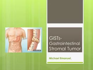
GISTs- Gastrointestinal Stromal Tumor. Michael Emanuel. Background. Representing 1-3% of gastrointestinal malignancies. The most common mesenchymal tumors of the gastrointestinal tract. Can arise anywhere along the gastrointestinal tract. Background Cont. Most GISTs are sporadic.

E N D
GISTs-Gastrointestinal Stromal Tumor Michael Emanuel.
Background • Representing 1-3% of gastrointestinal malignancies. • The most common mesenchymal tumors of the gastrointestinal tract. • Can arise anywhere along the gastrointestinal tract.
Background Cont. • Most GISTs are sporadic. • Arise from a common precursor cell. • Most commonly resulting from activating mutations in one of the receptor protein tyrosine kinases.
Epidemiology. • Occur in 10-20 per one million people. • Present at ages 50–70 years. • Similar in men and women. • Most GISTs are sporadic.
Mesenchymal tumors. • Mesenchymal tissue neoplasms are soft tissue tumors, also known as connective tissue tumors (bone, cartilage, fat, muscle,nerve, vascular, or hematopoietic tissues). • sarcomais a cancer that arises from transformed cells of mesenchymal origin. • Gists are the most common mesenchymal tumors of the gastrointestinal tract.
Presentation. Can arise anywhere along the gastrointestinal tract: • Stomach (50%). • Small intestine (25%). • Colon (10%). • Omentum/mesentery (7%). • Esophagus (5%). • Rectum (3%).
Signs and symptoms. Depending on the anatomic location of the tumor and the tumor size and aggressiveness: • GI bleeding. • An acute abdomen caused by tumor rupture. • Dysphagia. • Satiety. • GI obstruction. • Fatigue. • Anorexia, Weight loss, Nausea.
Pathophysiology(1). The neoplastic GIST cells appear to arise from a common precursor cell, which gives rise to the interstitial cells of Cajal in the normal myenteric plexus.
Pathophysiology(2). The discovery of gain-of-function mutantions in the c-Kit/PDGFRA proto-oncogene in GISTs that allowed GISTs to be distinguished reliably from these other histopathological subtypes of GI mesencymal tumors.
Characteristics(1). GISTs range in size from incidental lesions a few millimeters in diameter to large masses of 35 cm or more. The median size at presentation is approximately 5 cm.
Characteristics(2). The tumors are generally centered on the bowel wall, but may form polypoidserosal- or mucosal based masses.
Characteristics(3). Most GISTs present as a single, well circumscribed nodule. Rarely, a patient will have 2 separate GISTs at different locations in the gastrointestinal tract.
Management of Patients With GIST. • include history and physical examination. • appropriate imaging of abdomen and pelvis. • Surgery, or surgery + Post-surgical adjuvant treatment, or neoadjuvant + surgery. • Patients presenting with an acute abdomen require immediate surgery.
Diagnosis. • CT- for initial evaluation and surveillance for recurrence. • EUS- determines size and extent of the tumor. • PET-CT- reveals small metastases, establish baseline metabolic activity and assess therapy response.
Diagnosis. • Biopsy- examines the histopathology to identify the characteristics of GISTs. • Immunohistochemistry- specific antibodies that stain the molecule CD117 [also known as c-kit]. 95% of all GISTs are CD117-positive. (other possible markers include CD34, DOG-1, desmin, and vimentin).
Histophatology. Most GISTs show 1 of 3 histologic patterns: • spindle cell type (70%). • predominantly epithelioidcell type (20%). • mixture of both spindle and epithelioidcells(10%).
Prognostic Factors. The most important and widely used prognostic features of a primary tumor—their size, site and mitotic index.
Treatment. • In localized, resectable adult GISTs, if anatomically and physiologically feasible, surgery is the primary treatment of choice. • Radiotherapy has not historically been effective for GISTs. • GISTs do not respond to most chemotherapy medications. • Biological therapyhas been identified for clinical benefit in GIST: imatinib.
Imatinib (Gleevec). • Imatinibmesylate is a selective, potent, small molecule inhibitor of a family of structurally related tyrosine kinase signaling enzymes. • Inhibits both c-kit tyrosine kinase mutations and PDGFRA mutations. • Has been used in neoadjuvant and adjuvant settings.
Mechanism of action. By occupying the ATP binding pocket of the ABL kinase domain, imatinib prevents substrate phosphorylation and downstream activation of signals.
References. • NCCN Task Force Report: Management of Patients with Gastrointestinal Stromal Tumor (GIST)—Update of the NCCN Clinical Practice Guidelines. • Mod Pathol 2003;16(4):366–375, Gastrointestinal Stromal Tumors and Other Mesenchymal Lesions of the Gut, Joel K GreensonM.D.1, 1Department of Pathology, University of Michigan Health System, Ann Arbor, Michigan. • Miettinen M, Lasota J (2006). "Gastrointestinal stromal tumors: review on morphology, molecular pathology, prognosis, and differential diagnosis". Arch Pathol Lab Med 130. • Gastrointestinal stromal tumors: Pathology and prognosis at different sites MarkkuMiettinen, MD the corresponding author, Jerzy Lasota, MD.