Download
1 / 30
300 likes | 323 Views
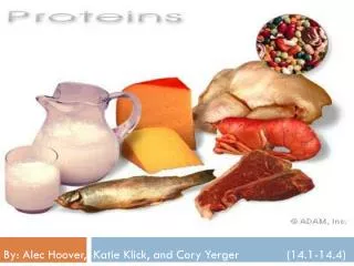
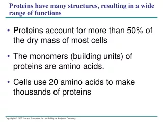
Proteins have many structures, resulting in a wide range of functions. Proteins account for more than 50% of the dry mass of most cells The monomers (building units) of proteins are amino acids. Cells use 20 amino acids to make thousands of proteins. Amino Acid Monomers.

E N D
Proteins have many structures, resulting in a wide range of functions • Proteins account for more than 50% of the dry mass of most cells • The monomers (building units) of proteins are amino acids. • Cells use 20 amino acids to make thousands of proteins
Amino Acid Monomers • Amino acids are organic molecules with carboxyl and amino groups a carbon Amino group Carboxyl group
Amino acids are linked by peptide bonds formed by dehydration reactions Peptide bond (a) Peptide bond Side chains Backbone Amino end (N-terminus) Carboxyl end (C-terminus) (b)
Polypeptides • Polypeptides are polymers of amino acids • A protein consists of one or more polypeptides Lysozyme: an antibacterial enzyme (protein) found in human tears. It is made of one polypeptide. Hemoglobin protein is made of four polypeptides
Fig. 5-22 Normal hemoglobin Sickle-cell hemoglobin Primary structure His Val Leu Glu Glu His Thr Val Primary structure Thr Pro Val Leu Pro Glu 1 2 3 4 5 6 7 1 2 3 4 5 6 7 Exposed hydrophobic region Secondary and tertiary structures Secondary and tertiary structures subunit subunit Sickle-cell hemoglobin Quaternary structure Normal hemoglobin (top view) Quaternary structure Function Molecules interact with one another and crystallize into a fiber; capacity to carry oxygen is greatly reduced. Function Molecules do not associate with one another; each carries oxygen. 10 µm 10 µm Red blood cell shape Normal red blood cells are full of individual hemoglobin moledules, each carrying oxygen. Red blood cell shape Fibers of abnormal hemoglobin deform red blood cell into sickle shape.
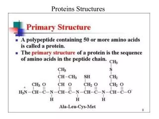
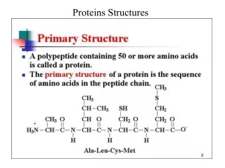
Four Levels of Protein Structure • Primary structure • Secondary structure • Tertiary structure • Quaternary structure
Primary structure, the sequence of amino acids in a protein, is like the order of letters in a long word
Secondary Structure • The coils and folds of secondary structure result from hydrogen bonds between atoms of the polypeptide backbone (NOT the amino acid side chain or R-groups). • Typical secondary structures are a coil called an alpha helix and a folded structure called a beta pleated sheet.
Fig. 5-21d Abdominal glands of the spider secrete silk fibers made of a structural protein containing -pleated sheets. The radiating strands, made of dry silk fibers, maintain the shape of the web. The spiral strands (capture strands) are elastic, stretching in response to wind, rain, and the touch of insects.
Tertiary Structure • Tertiary structure is determined by interactions between R groups, rather than interactions between backbone constituents • These interactions between R groups include: • hydrogen bonds, • ionic bonds, • hydrophobic interactions, • van der Waals interactions • Strong covalent bonds called disulfide bridges may reinforce the protein’s conformation.
Quaternary structure • Quaternary structure results when two or more polypeptide chains form one macromolecule • Collagen is a fibrous protein consisting of three polypeptides coiled like a rope • Hemoglobin is a globular protein consisting of four polypeptides: two alpha and two beta chains
Concept 5.5: Nucleic acids store and transmit hereditary information • The amino acid sequence of a polypeptide is programmed by a unit of inheritance called a gene • Genes are made of DNA, a nucleic acid
The Roles of Nucleic Acids • There are two types of nucleic acids: • Deoxyribonucleic acid (DNA) • Ribonucleic acid (RNA) • DNA replicates in order for the cells to divide • DNA directs the synthesis of messenger RNA (mRNA) and, through mRNA, controls protein synthesis • Protein synthesis occurs in ribosomes
Concept 3.1 Nucleic Acids Are Informational Macromolecules DNA’s information is encoded in the sequence of bases. DNA has two functions: Replication Information is copied to RNA and used to specify amino acid sequences in proteins.
DNA directs the synthesis of messenger RNA (mRNA) and, through mRNA, controls protein synthesis LE 5-25 DNA Synthesis of mRNA in the nucleus mRNA NUCLEUS CYTOPLASM mRNA Movement of mRNA into cytoplasm via nuclear pore Ribosome Synthesis of protein Amino acids Polypeptide
The Structure of Nucleic Acids • Nucleic acids are polymers called polynucleotides • Each polynucleotide is made of monomers called nucleotides • Each nucleotide consists of: • a nitrogenous base • a pentose sugar • and a phosphate group
LE 5-26a 5¢ end Nucleoside Nitrogenous base Phosphate group Pentose sugar Nucleotide 3¢ end Polynucleotide, or nucleic acid
Nitrogenous bases • There are two families of nitrogenous bases: • Pyrimidines have a single six-membered ring • Purines have a six-membered ring fused to a five-membered ring. Pyrimidines Nitrogenous bases Cytosine C Thymine (in DNA) T Uracil (in RNA) U Purines Adenine A Guanine G
Pentose sugar • In DNA, the sugar is deoxyribose • In RNA, the sugar is ribose. Pentose sugars Deoxyribose (in DNA) Ribose (in RNA)
The DNA Double Helix • A DNA molecule has two polynucleotides spiraling around an imaginary axis, forming a double helix • The nitrogenous bases in DNA form hydrogen bonds in a complementary fashion: A always pairs with T, and G always pairs with C
Concept 3.1 Nucleic Acids Are Informational Macromolecules The two strands are antiparallel (running in opposite directions), and the double helix is right-handed.
RNA • RNA is made of one polynucleotide (one strand of nucleotides) • The nucleotide of RNA is made of: • A nitrogenous bases, Adenine (A), Uracil (U), Guanine (G), or Cytosine (C). • A ribose sugar. • A phosphate group.
DNA replication and transcription depend on base pairing: 5′-TCAGCA-3′ 3′-AGTCGT-5′ transcribes to RNA with the sequence 5′-UCAGCA-3′. Concept 3.1 Nucleic Acids Are Informational Macromolecules