Download
1 / 49
600 likes | 1.05k Views
CAPNOGRAPHY In Emergency Care. EDUCATIONAL SERIES. Part 2: Introduction. Part 2: Introduction to Capnography. CAPNOGRAPHY In Emergency Care. Part 2: Introduction to Capnography Learning Objectives. Differentiate between oxygenation and ventilation Define end-tidal CO 2

E N D
CAPNOGRAPHYIn Emergency Care EDUCATIONAL SERIES Part 2:Introduction
Part 2: Introduction to Capnography CAPNOGRAPHYIn Emergency Care
Part 2: Introduction to Capnography Learning Objectives • Differentiate between oxygenation and ventilation • Define end-tidal CO2 • Identify phases of a normal capnogram • Recognize patterns of hypoventilation, hyperventilation and bronchospasm
Oxygenation and Ventilation What is the difference?
Oxygenation and Ventilation • Two completely different and separate functions • Oxygenation is the transport of O2 via the bloodstream to the cells • Oxygen is required for metabolism • Ventilation is the exhaling of CO2 via the respiratory tract • Carbon dioxide is a byproduct of metabolism
Oxygenation and Ventilation Ventilation (capnography) Oxygenation (oximetry) O2 Cellular Metabolism CO2
Oxygenation • Measured by pulse oximetry (SpO2) • Noninvasive measurement • Percentage of oxygen in red blood cells • Changes in ventilation take minutes to be detected • Affected by motion artifact, poor perfusion and some dysrhythmias
Oxygenation Pulse Oximetry Sensors Pulse Oximetry Waveform
Ventilation • Measured by the end-tidal CO2 • Partial pressure (mmHg) or volume (% vol) of CO2 in the airway at the end of exhalation • Breath-to-breath measurement provides information within seconds • Not affected by motion artifact, poor perfusion or dysrhythmias
Ventilation Capnography Lines Capnography waveform
Oxygenation versus Ventilation • Monitor your own SpO2 and EtCO2 • SpO2 waveform is in the second channel • EtCO2 waveform is in the third channel
OxygenationversusVentilation • Now hold your breath • Note what happens to the two waveforms SpO2 EtCO2 How long did it take the EtCO2 waveform to go flat line? How long did it take the SpO2 to drop below 90%?
Oxygenation Oxygen for metabolism SpO2 measures % of O2 in RBC Reflects change in oxygenation within 5 minutes Ventilation Carbon dioxide from metabolism EtCO2 measures exhaled CO2 at point of exit Reflects change in ventilation within 10 seconds Oxygenation and Ventilation
Why Measure Ventilation—Intubated Patients • Verify and document ET tube placement • Immediately detect changes in ET tube position • Assess effectiveness of chest compressions • Earliest indication of ROSC • Indicator of probability of successful resuscitation • Optimally adjust manual ventilations in patients sensitive to changes in CO2
Why Measure Ventilation—Non-Intubated Patients • Objectively assess acute respiratory disorders • Asthma • COPD • Possibly gauge response to treatment
Why Measure Ventilation—Non-intubated Patients • Gauge severity of hypoventilation states • Drug and ETOH intoxication • Congestive heart failure • Sedation and analgesia • Stroke • Head injury • Assess perfusion status • Noninvasive monitoring of patients in DKA
Interpreting EtCO2 and the Capnography Waveform • Interpreting EtCO2 • Measuring • Physiology • Capnography waveform
A r t e r y V e i n O x y g e n O 2 C O 2 O 2 End-tidal CO2 (EtCO2) Pulmonary Blood Flow Ventilation Left Atrium Right Ventricle Perfusion
End-tidal CO2 (EtCO2) • Carbon dioxide can be measured • Arterial blood gas is PaCO2 • Normal range: 35-45mmHg • Mixed venous blood gas PeCO2 • Normal range: 46-48mmHg • Exhaled carbon dioxide is EtCO2 • Normal range: 35-45mmHg
a-A Gradient Arterial to Alveolar Difference for CO2 Ventilation Left Atrium Right Ventricle Alveolus A r t e r y V e i n EtCO2 PaCO2 Perfusion
End-tidal CO2 (EtCO2) • Normal a-A gradient • 2-5mmHg difference between the EtCO2and PaCO2 in a patient with healthy lungs • Wider differences found • In abnormal perfusion and ventilation • Incomplete alveolar emptying • Poor sampling
End-tidal CO2 (EtCO2) • Reflects changes in • Ventilation- movement of air in and out of the lungs • Diffusion- exchange of gases between the air-filled alveoli and the pulmonary circulation • Perfusion - circulation of blood
End-tidal CO2 (EtCO2) • Monitors changes in • Ventilation- asthma, COPD, airway edema, foreign body, stroke • Diffusion - pulmonary edema, alveolar damage, CO poisoning, smoke inhalation • Perfusion- shock, pulmonary embolus, cardiac arrest, severe dysrhythmias
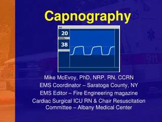
Capnographic Waveform • Normal waveform of one respiratory cycle • Similar to ECG • Height shows amount of CO2 • Length depicts time
Capnographic Waveform • Waveforms on screen and printout may differ in duration • On-screen capnography waveform is condensed to provide adequate information the in 4-second view • Printouts are in real-time • Observe RR on device
Capnographic Waveform • Capnograph detects only CO2from ventilation • No CO2 present during inspiration • Baseline is normally zero Baseline
Capnogram Phase IDead Space Ventilation • Beginning of exhalation • No CO2 present • Air from trachea, posterior pharynx, mouth and nose • No gas exchange occurs there • Called “dead space”
Capnogram Phase I Baseline B A I Baseline Beginning of exhalation
Capnogram Phase IIAscending Phase • CO2from the alveoli begins to reach the upper airway and mix with the dead space air • Causes a rapid rise in the amount of CO2 • CO2now present and detected in exhaled air
Capnogram Phase IIAscending Phase C Ascending Phase Early Exhalation II B A CO2 present and increasing in exhaled air
Capnogram Phase IIIAlveolar Plateau • CO2 rich alveolar gas now constitutes the majority of the exhaled air • Uniform concentration of CO2from alveoli to nose/mouth
C D I I I A B Capnogram Phase IIIAlveolar Plateau CO2 exhalation wave plateaus Alveolar Plateau
Capnogram Phase IIIEnd-Tidal • End of exhalation contains the highest concentration of CO2 • The “end-tidal CO2” • The number seen on your monitor • Normal EtCO2 is 35-45mmHg
Capnogram Phase IIIEnd-Tidal End of the the wave of exhalation D C End-tidal A B
Capnogram Phase IVDescending Phase • Inhalation begins • Oxygen fills airway • CO2 level quickly drops to zero
Capnogram Phase IVDescending Phase Inspiratory downstroke returns to baseline C D Descending Phase Inhalation I V A B E
Capnography Waveform Normal range is 35-45mm Hg (5% vol) Normal Waveform
Capnography Waveform Question • How would your capnogram change if you intentionally started to breathe at a rate of 30? • Frequency • Duration • Height • Shape
Hyperventilation RR : EtCO2 Normal Hyperventilation 4 5 0
Capnography Waveform Question • How would your capnogram change if you intentionally decreased your respiratory rate to 8? • Frequency • Duration • Height • Shape
4 5 0 4 5 0 Hypoventilation RR : EtCO2 Normal Hypoventilation
Normal Hyperventilation 4 5 0 Hypoventilation 4 5 0 Capnography Waveform Patterns
Capnography Waveform Question How would the waveform shape change during an asthma attack?
Bronchospasm Waveform Pattern • Bronchospasm hampers ventilation • Alveoli unevenly filled on inspiration • Empty asynchronously during expiration • Asynchronous air flow on exhalation dilutes exhaled CO2 • Altersthe ascending phase and plateau • Slower rise inCO2 concentration • Characteristic pattern for bronchospasm • “Shark Fin” shape to waveform
4 5 0 Capnography Waveform Patterns Normal Bronchospasm
Part 2: Introduction to Capnography Summary • Oxygenation and ventilation • Pulse oximetry • Measures O2 saturation in blood • Slow to indicate change in ventilation • Capnography • Measures CO2 in the the airway • Provides a breath-to-breath status of ventilation
Part 2: Introduction to Capnography Summary • Capnographic waveform has four phases • The highest CO2 concentration is at the end of alveolar plateau • End-tidal CO2 • Normal EtCO2 range is 35-45mmHg • Several conditions can be immediately detected with capnography
4 5 0 4 5 0 4 5 0 Capnography Waveform Patterns Normal Hyperventilation Hypoventilation Bronchospasm
Part 2: Introduction to Capnography We’re off to a running start!