Download
1 / 94
960 likes | 1.06k Views
Learn how alkylating agents induce cell death by forming covalent bonds with DNA and other biomolecules. Explore the mechanisms and resistance of cancer cells to these drugs.

E N D
Pharmaceutical chemistry Assist . Prof . Karima F. Ali Al-mustansiriyah university college of pharmacy Antineoplastic agents
Anticancer drugs The ability of drugs to kill cancer cells is generally believed to be because of their ability to induce the process of apoptosis. In high-dose therapy, cell death may occur by necrosis but this is also toxic to the patient. In a general sense the anti neoplastics target DNA or the process of DNA replication and stimulate apoptosis but the exact mechanisms by which this stimulation occurs are not known with certainty. The effectiveness of the agents is reduced in cells where apoptosis fails to occur properly, and this is a property of many cancer cells. Normal cells with fully functioning apoptotic mechanisms may then become susceptible to the action of the anti neoplastics increasing the toxicity of the agents.
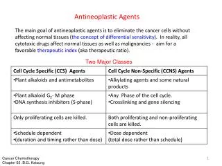
Drug classes 1.Alkylating Agents: The alkylating agents are a class of drugs that are capable of forming covalent bonds with important biomolecules. The major targets of drug action are nucleophilic groups present on DNA (especially the 7-position of guanine); however, proteins and RNA among others may also be alkylated.
Alkylation of DNA is thought to lead to cell death, although the exact mechanism is uncertain. Potential mechanisms of cell death include activation of apoptosis caused by p53 activation and disruption of the template function of DNA. The cancer cells have dysfunctional p53 so that even though the cell has been unable to replicate DNA error free, cell death via apoptosis does not occur. Cancer cells may become resistant to the effects of alkylating agents.
There are several potential nucleophilic sites on DNA, which are susceptible to electrophilic attack by an alkylating agent (N-2, N-3, and N-7 of guanine, N-1, N-3, and N-7 of adenine, 0–6 of thymine, N-3 of cytosine). The most important of these for many alkylating agents is the N-7 position of guanine whose nucleophilicity may be enhanced by adjacent guanine residues.
The general mechanism for alkylation involves nucleophilic attack by –N=, -NH2, -OH, -O-PO3H of DNA and RNA, while additional nucleophiles (-SH, COOH, etc.) present on proteins may also react.
Nitrogen mustards Mustards such as mechlorethamine are classified as dialkylating agents in that one mustard molecule can alkylate two nucleophiles. The initial acid–base reaction is necessary to release the lone pair of electrons on nitrogen, which subsequently displaces chloride to give the highly reactive aziridinium cation. Nucleophilic attack can then occur at the aziridinium carbon to relieve the small ring strain and neutralize the charge on nitrogen.
Mechlorethamine is highly reactive, in fact, too reactive and therefore nonselective, making it unsuitable for oral administration and necessitating direct injection into the tumor. In cases of extravasation (drug escapes from the tumor into the underlying tissue), the antidote sodium thiosulfate (Na2S2O3), a strong nucleophile, may be administered.
Chlorambucil and Melphalan The lack of selectivity of mechlorethamine led to attempts to improve on the agent. One rationale was to reduce the reactivity by reducing the nucleophilicity of nitrogen, thereby slowing aziridinium cation formation. This could be accomplished by replacement of the weakly electron-donating methyl group with groups that were electron withdrawing (-I). This is seen in the case of chlorambucil and melphalan by Attachment of nitrogen to a phenyl ring.
Reactivity was reduced such that these compounds could be administered orally. In the case of melphalan, attachment of the mustard functionality to a phenylalanine moiety was not only an attempt to reduce reactivity but also an attempt to increase entry into cancer cells by utilization of carrier mediated uptake. Melphalan was found to utilize active transport to gain entry into cells, but selective uptake by cancer cells has not been demonstrated
Cyclophosphamide and Ifosfamide Attachment of more highly electron-withdrawing functionalities was utilized in the case of cyclophosphamide and ifosfamide. In these cases, aziridinium cation formation is not possible until the electron-withdrawing function has been altered.
The drug could be selectively activated in cancer cells because they were believed to contain high levels of phosphoramidase enzymes. This would remove the electron-withdrawing phosphoryl function and allow aziridine formation to occur. The drug was activated by cytochrome P450 (CYP) isozymes CYP2B6 and CYP3A4/5 to give a carbinolamine that could undergo ring opening to give the aldehyde.
The increased acidity of the aldehyde -hydrogen facilitates a retro-Michael decomposition The ionized phosphoramide is now electron-releasing via induction and allows aziridinium cation formation to proceed. To decrease the incidence of kidney and bladder toxicity, the sulfhydryl (MSH) containing agent mesna may be administered and functions to react with the electrophilic species that may be present in the kidney.
there are differences in the metabolism and activity of the agents. Both are administered as racemic mixtures as a result of the presence of a chiral phosphorus atom. There appears to be little difference in the metabolic fate of the R- and S-isomers of cyclophosphamide, but in the case of ifosfamide, the R-isomer is converted to the required 4-hydroxy-ifosfamide 2 to 3 times faster than the S-isomer..
The S-isomer undergoes preferential oxidation of the side chain to give N-dechloroethylation, which removes the ability of the agent to cross-link DNA and also produces the neurotoxic and urotoxic chloroacetaldehyde. An additional difference between cyclophosphamide and ifosfamide is the larger alkylating species that ultimately results after metabolic activation of ifosfamide. This results in the reactive form of ifosfamide having a higher affinity for DNA than the analogous form of cyclophosphamide and differences in the interstrand and intrastrand links that ultimately result.
Organoplatinum compounds There are several organometallic compounds based on platinum that play a central role in many cancer treatment protocols. The first of these, cisplatin. Less reactiveplatinum compounds such as carboplatin and oxaliplatin in which the leaving group was incorporated into a chelate.satraplatin is currently in clinical trials. One advantage of these agents is the possibility of oral administration. Satraplatin has shown similar activity when given orally to that of cisplatin given by injection.
Mechanism of cisplatin activation and formation of DNA adducts.
Nitrosoureas Compound was based on the idea that its chemical decomposition was leading to the formation of diazomethane (CH2N2) and subsequent alkylation of DNA, this led to the nitrosoureas, where it was found that activity could be enhanced by attachment of a 2-haloethyl substituent to both nitrogens
These compounds are reasonably stable at pH 4.5 but undergo both acid and base catalyzed decomposition at lower and higher pH, respectively. There are several pathways of decomposition that are possible for these compounds, but the one that appears to be most important for alkylation of DNA involves: Abstraction of the NH proton, which is relatively acidic (pKa 8–9). Rearrangement to give an isocyanate and a diazohydroxide.
The diazohydroxide, upon protonation followed by loss of water, yields a diazo species that decomposes to a reactive carbocation. The isocyanate functions to carbamylate proteins and RNA, whereas the carbocation is believed to be the agent responsible for DNA alkylation.
Detoxification pathways of the nitrosoureas are also possible and can play a role in resistance to this group of agents. The first of these involves dechlorination, which is facilitated by CYP participation. The second route involves denitrosation,
2.Antimetabolites Most antimetabolites are effective cancer chemotherapeutic agents via interaction with the biosynthesis of nucleic acids. Therefore, several of the useful drugs used in antimetabolite therapy are purines, pyrimidines, folates, and related compounds. The antimetabolite drugs may exert their effects by several individual mechanisms involving enzyme inhibition at active, allosteric, or related sites.
The purine and pyrimidine antimetabolites are often compounds incorporated into nucleic acids and the nucleic acid polymers (DNA, RNA, etc.). • The antifolates are compounds designed to interact at cofactor sites for enzymes involved in the biosynthesis of nucleic acid bases.
A. Pyrimidine Drugs The pyrimidine derivative 5-fluorouracil (5-FU) was designed to block the conversion of uridine to thymidine. The normal biosynthesis of thymidine involves methylation of the 5-position of the pyrimidine ring of uridine. The replacement of the hydrogen at the 5-position of uracil with a fluorine results in an antimetabolite drug, leading to the formation of a stable covalent ternary complex composed of 5-FU, thymidylate synthase (TS), and cofactor (a tetrahydrofolate species).
The metabolic activation (anabolism) of 5-FU required to produce the anticancer effects accounts for no more than 20% of the administered amount of drug in most patients. Catabolic inactivation via the normal pathways for uracil consumes the remaining approximate 80% of the dose. The major enzyme of pyrimidine catabolism is dihydropyrimidinedehydrogenase (DPD), and 5-FU is a substrate for this enzyme.
Uracil is a substrate for this enzyme system also and has been dosed with 5-FU and 5-FU prodrugs in an attempt to saturate DPD and conserve active drug species. Variability in the levels of DPD activity among the patient population is a major factor in the bioavailability of 5-FU. low bioavailability of 5-FU as a result of the catabolic efficiency of DPD and other enzymes has lead to the development of unique dosing routes and schedules as well as the development of prodrug forms of 5-FU
TS is responsible for the reductive methylation of deoxyuridine monophosphate (dUMP) by 5,10-methylenetetrahydrofolate to yield dTMP and dihydrofolate. Because thymine is unique to DNA, the TS enzyme system plays an important role in replication and cell division. • The tetrahydrofolate cofactor species serves as both the one-carbon donor and the hydride source in this system.
The initial step of the process involves the nucleophilic attack by sulfhydryl group of a cystine residue at the 6-position of dUMP. The resulting enolate adds to the methylene of 5,10- CH2-THF perhaps activated via the very reactive N-5- iminium ion .The iminium ion likely forms at N-5 and only after 5,10-CH2-THF binds to TS. The iminium ion is likely formed at N-5 because it is the more basic of the two nitrogens, whereas N-10 is the better leaving group.
The loss of the proton at the 5-position of dUMP and elimination of folate yields the exocyclic methylene uracil species. The final step involves hydride transfer from THF and elimination to yield the enzyme, DHF, and dTMP. • Attempts at chemical modification of 5-FU to protect from catabolic events have produced several prodrug forms, which are converted via in vivo metabolic and/or chemical transformation to the parent drug 5-FU. • The carbamate derivative of 5-deoxy-5-fluorocytidine is known as capecitabine, and it is converted to 5-FU through a series of activation steps.
The tetrahydrofuran derivative tegafur is slowly converted to 5-FU but requires quite high doses to reach therapeutic plasma concentrations. • Esters of the N-hydroxymethyl derivative of tegafur show greater anticancer activity than tegafur.
5-Fluorouracil is activated by conversion to the corresponding nucleotide species, 5-fluoro-2-deoxyuridylic acid. The resulting 5-fluoro-2-deoxyuridylic acid is a powerful inhibitor of thymidylate synthetase , the enzyme that converts 2-deoxyuridylic acid to thymidylic acid.
In the inhibiting reaction, the sulfhydryl group of TS adds via conjugate addition to the 6-position of the fluorouracil moiety. The carbon at the 5-position then binds to the methylene group of 5,10-Methylene tetrahydrofolate following initial formation of the more electrophilic form of folate the N-5-iminium ion. In the normal process, this step is followed by the elimination of dihydrofolate from the ternary complex, regeneration of the active enzyme species, and the product thymidine.
Central to this process is the loss of the proton at the 5- position of uracil to form the exocyclic methylene uracil species. The 5-fluorine is stable to elimination, and a terminal product results, involving the enzyme, cofactor, and substrate, all covalently bonded.
Cytarabine and gemcitabine Pyrimidine analogs as antimetabolites for cancer therapy have been developed based on the cytosine structure as well. Modification of the normal ribose or deoxyribose moiety has produced useful drug species such as cytarabine (ara-C) and gemcitabine. Cytosine arabinoside (ara-C or cytarabine) is simply the arabinose sugar instead of ribose, and the only difference in structure is the epimeric hydroxyl group at the 2-position of the pentose sugar.
Mechanism of action may include a slowing of the DNA chain elongation reaction via DNA polymerase or cellular inefficiencies in DNA processing or repair after incorporation. Gemcitabine is the result of fluorination of the 2`- position of the sugar moiety. After its anabolism to diphosphate and triphosphate metabolites, it inhibits ribonucleotide reductase and competes with 2-deoxycytidine triphosphate for incorporation into DNA. The mechanism of action for gemcitabine is likely similar to that of ara-C including alteration of the rate of incorporation into DNA as well as the rate of DNA processing and repair.
Modification of the pyrimidine ring has also been explored for the development of potential anticancer drugs based on antimetabolite theory. Several pyrimidine nucleoside analogs have one more or one less nitrogen in the heterocyclic ring. They are known as azapyrimidine or deazapyrimidine nucleosides. 5-Azacytidine is an example of a drug in this category The mode of action of this compound is complex involving reversible inhibition of DNA methyl transferase, and this lack of methylated DNA activates tumor suppressor genes. In certain tumor systems, it is incorporated into nucleic acids, which may result in misreading or processing errors.