Download

1 / 25
250 likes | 417 Views
Extraction of Human DNA. Experiment Goals. Isolation of genomic DNA from human blood Analysis of isolated DNA using Agarose gel electrophoresis Spectrophotometry. What is a DNA?. DNA, also known as deoxyribonucleic acid, A fundamental molecule found in all living things

E N D
Experiment Goals • Isolation of genomic DNA from human blood • Analysis of isolated DNA using • Agarose gel electrophoresis • Spectrophotometry
What is a DNA? • DNA, also known as deoxyribonucleic acid, • A fundamental molecule found in all living things • Carries the genetic information in the cell • Contains instructions for our body cells to perform their specific functions • The sequence of nucleotides determines individual hereditary characteristics
What is a DNA? • Basic unit of information in DNA is the gene • Human beings have about 30,000 gene • Size of organism’s genome is roughly a measure of its complexity • Viruses 5-10 kb • E. coli 4,640 kb • Human 2,900,000 kb
DNA Extraction • DNA extraction is a routine procedure to isolate & collect DNA. • DNA extraction is the first step for subsequent molecular or forensic analysis. • DNA can be extracted from almost any intact cellular tissue • Skin, • blood, • saliva, • semen, • mucus, • muscle tissue, • bone marrow, etc.
Nucleic Acid Preparation Applications • Medical studies • Understanding genetic disorders at molecular level • Rapid detection of genetic disorders in a patient • Agricultural studies • Plant and animal breeding • Criminology/Paternity testing • DNA fingerprinting to identify individuals.
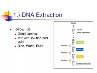
Basic steps in DNA extraction • There are three basic steps in a DNA extraction, the details of which may vary depending on the type of sample and any substances that may interfere with the extraction and subsequent analysis. • Break open cells and remove membrane lipids • Remove cellular and histone proteins bound to the DNA, by adding a protease, by precipitation with sodium or ammonium acetate, or by using a phenol/chloroform extraction step. • Precipitate DNA in cold ethanol or isopropanol, DNA is insoluble in alcohol and clings together, this step also removes salts.
1- Lyse RBCs & WBCs 2- Lyse WBCs nuclei & Denature/digest proteins 3- Separate contaminants (e.g., proteins, heme) 4- Precipitate DNA 5- Resuspend DNA in final buffer Overview of Procedure
Blood Collection • Blood collected in disodium EDTA tube • Samples can be stored at -20oC or -70oC • Fresh samples are kept in freezer for a few hours to facilitate RBCs hemolysis • Allow samples to thaw before starting the extraction
1- RBCs Lysis • Pipette 3 mls of whole blood in a conical centrifuge tube • Add 9 mls of 1X erythrocyte lysing buffer(0.155M NH4Cl, 10 mM KHCO3, 0.1 mM Na2EDTA, pH 7.4) • Leave 10 min. at RT, mix occasionally • Centrifuge at 4000 rpm for 5 min • Discard supernatent • White pellet is observed at bottom of tube • Wash pellet 3 times by adding 3 mls of buffer, incubate 10 min at RT, & centrifuge
2- WBCs nuclei Lysis & proteins digestion • Add 1.5 mls of SE buffer (75mM NaCl, 25 mM Na2EDTA, pH 8.0) containing 100µg/ml of Proteinase K & 1% sodium dodecyl sulphate (SDS) to the pellet • Incubate at 37-55oC overnight in a water bath or incubator • WBCs nuclei denatured & DNA goes out in solution
3- Separate contaminants from DNA • After incubation add 1.5 mls of SE buffer, 750 µl of 6M NaCl & 3.75 mls chloroform • Mix vigorously on vortex for 20 sec • Mix for 30 min (on rotator) • Centrifuge for 10 min at 2000 rpm • 2 phases are observed • DNA is extracted in supernatant & proteins in the lower phase • Transfer upper aqueous phase (containing DNA) to a clean tube
4- Precipitate DNA • Add an equal volume of isopropanol • DNA will be precipitated by gentle swirling & observed as a white thread like strand • Using a sterile spatula or loop transfer the DNA strand into a sterile microcentrifuge tube containing 1 ml of 75% ethanol • Wash by inversion to remove any remaining salts • Centrifuge, discard supernatent • Repeat the washing step, then centrifuge • Remove supernatant, and dry the pellet
5- Resuspend DNA in final buffer • Dried pellet is resuspended in TE buffer and left overnight on a rotator
DNA Analysis • Different methods for assessing quantity & quality of extracted DNA • Agarose gel electrophoresis • UV spectrophotometry
Checking the Quality of DNA • The product of DNA extracted will be used in subsequent experiments • Poor quality DNA will not perform well in PCR
Quality from Agarose Gel Electrophoresis • Quality of DNA extracted is assessed using the following simple protocol: • Mix 5 µL of DNA with 5 µL of loading Dye • Load this mixture into a 1% agarose gel • Stain with ethidium bromide • Electrophorese at 70–80 volts, 45–90 minutes.
DNA Quality fromAgarose Gel Electrophoresis • High molecular weight band • Smearing indicates DNA degradation
Nucleic Acid Characterization • Absorption Spectra • Absorb light in ultraviolet range, most strongly in the 254-260 nm range • Useful for quantification of samples
Multiply the concentration of the DNA sample by the volume of hydrating solution added. Example for DNA: 150 µg/mL X 0.1 mL = 15 µg Concentration from UV Spec. (µg DNA per ml of hydrating solution) Volume of hydration solution DNA yield Quantity from UV Spectrophotometry Calculating Yield
Purity from UV Spectrophotometry • DNA absorb maximally at 260 nm. • Proteins absorb at 280 nm. • Background scatter absorbs at 320 nm.
A260/A280 = measure of purity (A260 – A320)/(A280 – A320) 1.7 – 2.0 = good DNA or RNA <1.7 = too much protein or other contaminant Purity from UV Spectrophotometry
<4 Months 1–3 Years <7 Years >7 Years 2–25 °C 2–8 °C –20 °C –70 °C Recommended for long-term storage in ethanol Storage Conditions • Store DNA in TE buffer at 4 °C for weeks or at –20 °C to –80 °C for long term.