Download
1 / 1
10 likes | 104 Views
This figure illustrates the gross anatomy of the harvested human sciatic nerve alongside the average sciatic nerve from the study. Different color-coded branches and their locations are specified, aiding in understanding nerve composition. Detailed cross-sections presented in Figures 3-7 further enhance comprehension. The study provides insights for ankle control using nerve-cuff electrodes.

E N D
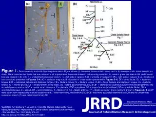
Figure 1. Gross anatomy and stick-figure representation. Figure shows (a) harvested human sciatic nerve next to (b) average sciatic nerve seen in our study. Black branches are those that are common to all 3 specimens. Branches shown in red are only present in 1L, ones in green are seen in 2R, and those in blue are present in 2L only. ? = unidentified cutaneous branch, 1L = left side of cadaver 1,2L = left side of cadaver 2, 2R = right side of cadaver 2, A = location of cross-sections presented in Figures 3–4, AD = adductor mag-nus, B = location of cross-section presented in Figures 5–6, DF = deep fibular, DL = digitorum longus, EXT = extensors hallucis and digitorum longus, FB = fibularis brevis, FL = fibularis longus, FLEX = flexor hallucis and digitorum longus, HL = hallucis longus, IG = inferior gluteal, LB = biceps femoris (long head), LG = lateral gastrocnemius, LSC/SCB* = lateral sural cutaneous/sural communicatingbranch, MG = medial gastrocnemius, MSC = medial sural cutaneous, P = plantaris, POP = popliteus, SB = biceps femoris (short head),SF = superficial fibular, SM = semimembranosus, S = soleus, ST = semitendinosus, T = tibial nerve, TA = tibialis anterior, TP = tibialis posterior. Cross-sections shown in Figures 3, 4, and 7 were taken from respectively marked locations on (b). *After harvesting, this branch (LSC/SCB) was more accurately identified as SCB and the unidentified cutaneous branch (?) was determined to be LSC. Gustafson KJ, Grinberg Y, Joseph S, Triolo RJ. Human distal sciatic nerve fascicular anatomy: Implications for ankle control using nerve-cuff electrodes. J Rehabil Res Dev. 2012;49(2):309–22.http://dx.doi.org/10.1682/JRRD.2010.10.0201