Chap 19 – Lymphatic System
310 likes | 1.13k Views
Chap 19 – Lymphatic System. Homework: Read Chap 19. Review anatomical structures & physiology (per diagrams). Learning Objectives: 1.List the functions of the lymphatic system. 2. Describe the source of lymph & its transport. 4. Describe lymph tissue.

Chap 19 – Lymphatic System
E N D
Presentation Transcript
Chap 19 – Lymphatic System Homework: Read Chap 19. Review anatomical structures & physiology (per diagrams). Learning Objectives: 1.List the functions of the lymphatic system. 2. Describe the source of lymph & its transport. 4. Describe lymph tissue. 5. Describe lymph nodes (locations structure and distribution of the lymph vessels. 3. Explain the, histology, function, etc.) 6. Name and describe other lymphoid organs of the body.
What’s the Problem? These patients have an imbalance called ___________.
Opening Discussion What does the lymphatic system do? How does it tie-in to what we have been studying? http://health.howstuffworks.com/adam-200102.htm
Circulatory Dilemma Problem: Capillaries are known to be ‘leaky’ vessels. They leak about 3L of interstitial fluid into tissue spaces daily. All this fluid must be returned to the cardiovascular system to ensure sufficient blood volume How does it get returned?
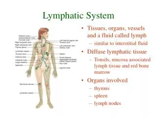
Lymphatic System To The Rescue! The lymphatic system - an elaborate system of drainage vessels that collect excess interstitial fluid and return it to the bloodstream
How Does It Work? Instructions: 1. Take time to reflect. 2. Review Figure 19.1 (page 680) and the associated text, “Distribution and Structure of Lymphatic Vessels”. 3. Write a 1-3 sentence summary in your own words of how it basically works. Place the summary in your notes next to this slide. 4. Be prepared to share with class. Estimated Time: 3-5 minutes
Lymph Vessels • A __________ system in which lymph flows toward the heart • Lymph vessels include: • Microscopic, permeable, blind-ended capillaries • Lymphatic collecting vessels • Trunks and ducts
Lymph Collecting Vessels • Have the same three ______ as veins • Have _______ walls, with more internal valves • Collecting vessels in the skin travel with superficial veins • Deep vessels travel with arteries
Lymph Capillaries • Similar to blood _________, with modifications • Remarkably ___________ • Loosely joined endothelial minivalves (withstand interstitial pressure and remain open). Also, function as one-way gates that: a) Allow interstitial fluid to enter lymph capillaries b) Do not allow lymph to escape from the capillaries
Lymph Capillaries continued • During inflammation, lymph capillaries can absorb: • _____ debris • __________ • ______ cells • Cells in the lymph nodes: • Cleanse and __________ this debris • Lacteals – specialized lymph capillaries present in intestinal mucosa • Absorb digested fat and deliver chyle (fatty lymph) to the blood
Lymph Transport • The lymphatic system _______ an organ that acts as a pump • Vessels are ____-pressure conduits • Uses the same methods as _____ to propel lymph • Pulsations of nearby arteries • Contractions of smooth muscle in the walls of the lymphatics
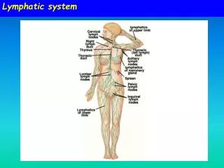
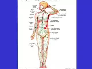
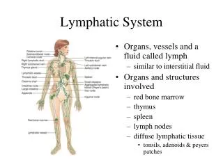
General Distribution of Vessels and Regional Lymph Nodes Label your practice diagram. Page 682
Lymphatic Trunks • Lymphatic trunks are formed by the union of the largest collecting ___________ • Major trunks include: • Paired ________, bronchomediastinal, subclavian, and ________ trunks • A single intestinal trunk • Lymph is delivered into one of two large ______ • _____ lymphatic duct – drains the right upper arm and the right side of the head and thorax • _______ duct – arises from the cisterna chyli and drains the rest of the body
Lymphatic Trunks Label your practice diagram Page 682
Lymphoid Cells • ____________ are the main cells involved in the immune response . The two main varieties are _ cells and _ cells (protect the body against foreign antigens such as bacteria, viruses, & cancer) • __________ – phagocytize foreign substances and help activate T cells • ________ cells – spiny-looking cells with functions similar to macrophages • ________ cells – fibroblastlike cells that produce a stroma, or network, that supports other cell types in lymphoid organs
Lymph Nodes • Lymph nodes are the principal lymphoid ________ of the body • Nodes are imbedded in _______ tissue and clustered along lymphatic vessels • Aggregations of these nodes occur near the body surface in inguinal, _______, and cervical regions of the body
Functions of Lymph Nodes • Their two basic functions are: • ____________ – macrophages destroy microorganisms and debris • ________ ______ __________ – monitor for antigens and mount an attack against them
Lymph Node Structure • ________ are bean shaped and surrounded by a fibrous capsule • Nodes are ______________ (trabeculae extend inward) • Nodes have two histologically distinct regions: a _______ and a _______
Lymph Node Structure continued Label your practice diagram.
Lymph Node Structure continued • The cortex contains _________ with germinal centers, heavy with dividing B cells • __ _______ circulate continuously among the blood, lymph nodes, and lymphatic stream • _________ cords extend from the cortex and contain B cells, T cells, and plasma cells • Throughout the node are lymph _______ crisscrossed by reticular fibers. Macrophages reside on these fibers and phagocytize ______ matter
Other Lymphoid Organs • The ____, ______ gland, and _______ • _________ patches and bits of lymphatic tissue scattered in connective tissue • All are composed of reticular connective tissue and all help protect the body • Only lymph nodes filter lymph
Spleen • _________ lymphoid organ, located on the left side of the abdominal cavity beneath the diaphragm • Functions • Site of lymphocyte proliferation • Immune surveillance and response • Cleanses the ______ • Stores blood ________
Spleen Structure, page 686 • Two distinct areas of the spleen are: • _____ pulp – area containing mostly lymphocytes suspended on reticular fibers and involved in immune functions • _____ pulp – remaining splenic tissue concerned with disposing of worn-out RBCs and bloodborne pathogens
Thymus • A bi-lobed organ that secrets hormones (thymosin and thymopoietin) that cause T lymphocytes to become immunocompetent • The size of the thymus varies with age • It increases in size and is most active during ________ • It stops growing during adolescence and then gradually _________
Thymus continued • The thymus _______ from other lymphoid organs in important ways • It functions strictly in T lymphocyte ________ • It does not directly fight _______ • The thymus consists of star-shaped epithelial cells (not reticular fibers)
Tonsils • Simplest lymphoid organs; form a ring of lymphatic tissue around the _______ • Like little crypts - ____ and ______ bacteria and particulate matter
Other continued • Peyer’s patches – isolated clusters of lymphoid tissue, similar to tonsils • Found in the wall of the distal portion of the _____ _________ • Similar structures are found in the appendix • _____ – mucosa-associated lymphatic tissue (protects the digestive and respiratory systems from foreign matter)