Download
1 / 56
730 likes | 1.77k Views
HEMOGLOBINOPATHIES: Thalassemia and Sickle Cell (with brief mention of a few others). Peter Newburger, MD Departments of Pediatrics and Cancer Biology. Epidemiology and the malaria hypothesis.

E N D
HEMOGLOBINOPATHIES:Thalassemia and Sickle Cell(with brief mention of a few others) Peter Newburger, MD Departments of Pediatrics and Cancer Biology
Epidemiology and the malaria hypothesis Distribution of thalassemias, sickle cell disease, G6PD mirror worldwide distribution of malaria prior to 20th century. Hypothesis (Haldane and others): heterozygous forms confer fitness. Thal trait, sickle trait, G6PD protective against death from cerebral falciparum malaria. Benefit outweighs homozygous risk. + o
ALPHA: Decreased or absent globin chains BETA: Decreased or absent globin chains ( DELTA and/or GAMMA thalassemia also possible, rare) SOME STRUCTURAL VARIANTS: The Thalassemia Syndromes
a and b chromosomal loci The molecular pathology of thalassemia is based on CHAIN IMBALANCE: In bthal, excess a In athal, too much b
αchains 100 βchains (adult) chains (fetal) 80 60 Globin chain percentage 40 εchains(embryonic) 20 δchains 0 0 -6 -3 Birth 3 6 Months Human Globin Synthesis DuringPrenatal and Neonatal Periods
Pathophysiology of Beta Thalassemia Normal production alpha chains Absent or decreasedbeta chains Unstable excess alpha tetramers Absent or decreased Hemoglobin A Hemolysis Dyserythropoiesis Weatherall. BMJ. 1997;314:1675.Olivieri et al. N Engl J Med. 1999;341:99.
Thalassemia Phenotypes: Based on Globin Chain Synthesis Alpha globin: Number of gene deletions • --/-- • -/-- • -/-,/-- • /- Beta globin: Level of gene expression • 0absent • + mod • + mild • + silent Weatherall. The Harvey Lectures. 2001;Series 94:1.
Beta Thalassemia: Clinical Heterogeneity Hb = hemoglobin; RBCs = red blood cells.
Phenotype Variability: Other Genetic Contributions Co-inheritance with beta-thal • Alpha gene deletions • Alpha globin enhancement (/, /) • Postnatal fetal hemoglobin production (HPFH) • Epigenetic modifiers (e.g. UDP-glucuronosyl-transferase gene, UGT1A1) Compound heterozygous states • Structural hemoglobinopathies (E, Lepore, Constant Spring) Weatherall. The Harvey Lectures. 2001;Series 94:1.
Peripheral Blood Smears • Thal major/intermedia • Severe hypochromia & microcytosis • Marked poikilocytosis • Target cells • Nucleated RBCs • Thal trait • Mild microcytosis • Mild or absent hypochromia • Fine basophilic stippling • Occasional targets
Differential diagnosis of microcytic anemia: thal trait, iron deficiency, or both
Differential diagnosis of microcytic anemia: thal trait, iron deficiency, or both Best diagnostic test: Therapeutic trial of iron Repeat H/H in 1 month (retic in 1 week if impatient)
INCREASED GIIRON ABSORPTION IRON OVERLOAD HEPATIC FIBROSIS/CIRRHOSISPANCREATITIS/DIABETESHYPOTHYROIDISM HYPOPITUITARISM HYPOPARATHYROIDISMOSTEOPOROSIS PULMONARY DYSFUNCTIONCARDIAC DISEASE–ARRHYTHMIAS MYOCARDOPATHY TRANSFUSIONS IRON CHELATION LATE DEATH Natural History of Thalassemia FAILURE TO THRIVECONGESTIVE HEART FAILURE TISSUE ANOXIA EARLY DEATH BONE MARROW EXPANSIONBONE DEFORMITIES INEFFECTIVE RBC PRODUCTION INTRAMEDULLARYEXTRAMEDULLARY ANEMIA HEPATOSPLENOMEGALYLYMPHADENOPATHY
Thalassemic bony changes Compression fracture “thalassemic facies” – Completely avoidable with modern transfusion therapy
Therapy • Chronic transfusions • Chelation therapy • Deferoxamine • Close follow-up for consequences of chelation therapy and iron overload
Iron Loading • Transfusions 200-250 mg iron per unit of RBC 5-6+ grams/yr for 50 kg patient • Abnormally regulated absorption 2-5 grams per year
Non-transferrin Bound Iron • Transferrin capacity exceeded • Non-transferrin bound iron in plasma promotes ˙OH radical formation • Peroxidative damage to membrane lipids/proteins • Myocyte damage • Mitochondrial respiratory chain impairment • Abnormal energy metabolism Annals of the New York Academy of Sciences 850:191-201 (1998)
Monitoring for Iron Overload • Ferritin • Very poor correlation with liver iron • Liver Biopsy • Confounders • Size of sample • Fibrosis and/or Cirrhosis • Superconducting Quantum Interference Device (SQUID) • New non-invasive measures • T2* cardiac MRI • R2 MRI of liver
Treatment: Chelation byDeferoxamine infusion • Subcutaneous administration • 5-7 days per week, 8-12 hrs • Tens of thousands of dollars per year • Reactions fairly common Blood, 2004, 104:34
New Chelation Drugs • Deferasirox(Exjade) • Newly licensed • Oral, once daily administration • Now the standard of care • Deferiprone (oral) • S-DFO –Starch deferoxamine compound (long-acting parenteral)
Improved Survival in Thalassemia • Historical poor prognosis • US Cohort born between 1960 and 1976: Median survival 17 years • Italian cohort born mid 1960s:Median survival to age 12 Haematologica 2004;89:1187-1193
Survival Curve by Birth Cohort 1.00 0.75 Survival Probability 0.50 P<0.00005 0.25 0.00 0 5 10 15 20 25 30 Age (years) Haematologica 2004;89:1187-1193
HSCT for Beta Thalassemia Major • First case report in 1982, Seattle • Subsequently >2300 transplants performed worldwide with >1000 in Pesaro, Italy • Most have received HLA-identical siblingallografts after myeloablative preparationwith busulfan/cyclophosphamide • Limited experience with alternate donors 1. Thomas et al. Lancet. 1982;2:227. 2. Sevilla et al. Bone Marrow Transplant. 2005;35:S17.
Event-Free Survival After HSCT for Beta Thalassemia, by Liver status Thalassemia-Free Survival in Patients Aged <17 y 1.0 Class 1 (n=146) 87% 0.8 84% Class 2 (n=334) 0.6 Probability 0.4 0.2 0 0 5 10 15 20 Years HLA-identical sibling donors, BU/CY conditioning Storb et al. Hematology (Am Soc Hematol Educ Program). 2003:372.
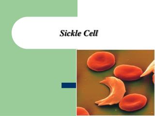
Hemoglobinopathies: Sickle and other abnormal globins
Hemoglobin name Beta globin gene mutation Amino acid substitution Hb Hijiyama 1341 A → G beta 120 Lys → Glu Hb Takamatsu 1341 A → C beta 120 Lys → Gln Hb Jianghua 1342 A → T beta 120 Lys → Ile Hb Riyadh 1343 A → C beta 120 Lys → Asn beta-thalassemia 1344 G → T beta 121 Glu → Stop Hb D-Los Angeles 1344 G → C beta 121 Glu → Gln Hb O-Arab 1344 G → A beta 121 Glu → Lys Hb St. Francis 1345 A → G beta 121 Glu → Gly Hb Beograd 1345 A → T beta 121 Glu → Val Hb D-Neath 1345 A → C beta 121 Glu → Ala Mutations affecting beta globin structure http://globin.cse.psu.edu/hbvar/menu.html
Sickle cell anemia From http://fig.cox.miami.edu/~cmallery/150/gene/mol_gen.htm
Sickle Cell Epidemiology bS : Single a.a. substitution Glutamic acid Valine Most common single gene disorder in African Americans 1/375 affected (homozygous) 1/12 are heterozygous carriers (~8%) Also affects other ethnicities: India, Middle East, Hispanic
Sickle Hemoglobinopathies • Beta globin Glu6Val: • Hemoglobin S insoluble • Long polymers break RBC membranes • Sickle partner hemoglobins • Hb C (Glu6Lys) • Hb D (several) • Hb OArab (Glu121Lys) • S-beta thalassemia (β0 vs. β+)
Genetic Modifiers of Severity in Sickle Cell Disease • Gamma globin: HPFH • Alpha globin: alpha thal trait • lower MCH (minimum gelling concentration) • lower MCV (viscosity) • UDP–glucuronosyltransferase 1A1 (UGT1A1) • Genes controlling inflammation • Genes controlling vascular adhesion • Genes controlling WBC?
Molecular Pathophysiology Intracellular [Hb] ~30 - 35 g/dL Deoxygenation allows interaction of bS subunits via abnormal hydrophobic regions (valines) Non-covalent bond with other bS in the RBC Formation of 14 stranded helical fiber
Polymerization phase is sensitive to: • pH (acidosis worse) • O2 concentration • Hemoglobin concentration • Ionic strength Bunn HF. NEJM 1997 337 (11)
Cellular Pathophysiology of Sickle Cell syndromes Polymerization leads to: Distortion of Cell shape Damage to RBC Membrane Abnormal permeability Irreversible sickling Impairment of RBC flow = Infarction Premature hemolysis = Anemia HbA and HbF inhibit polymerization of HbS by interference with the hydrophobic association.
Newborn Screening for Hb S 1987: NIH recommends screening for HbS be mandated by state law for every newborn. 2008: Universal Screening: 50 States plus D.C., U.S.V.I. & Puerto Rico Information on all newborn screening: http://genes-r-us.uthscsa.edu/
Hemoglobin Screening Nomenclature Hemoglobins are reported in order of quantity i.e. “FSA” = HbF > HbS > HbA FSA ≠ FAS EXAMPLES: • FA normal • FS Sickle Cell S/S (or S/β0 thalassemia) • FSC Hemoglobin SC disease • F Beta thalassemia major • FA(Barts) alpha thal trait or Hgb H (may also detect silent carriers) Trivial variants • Examples: Hb G, Hb Hasharon Imprecise identification: E/O, D/G
Sickle complications • Vaso-occlusive crisis • Lifelong chronic anemia • Cerebrovascular disease • Progressive vasculopathy • Ischemic stroke • Moyamoya disease and hemorrhagic stroke • Aplastic crisis • Splenic sequestration • Sepsis due to functional asplenia • Acute chest syndrome
Anemia • Most common feature of sickle cell disease • Often ignored as pathologic • Moderate to severe in almost all patients with SCD-SS (Hb 6-8.5 g/dL) Chronic intravascular hemolytic anemia • Degree of anemia reflects clinical severity • Episodic acute anemia • Red cell aplasia (parvovirus B19) • Acute splenic sequestration • Infection-related acute hemolysis
Sickle cell manifestations ACUTE CHRONIC • Vaso-occlusive Dactylitis / Hand Foot Syndrome CV: cardiomegaly, murmur Hepatosplenic sequestration Renal: isosthenuria, hematuria Priapism Eyes: proliferative retinopathy Pain crises (majority are bone) Skin: leg ulcers Acute chest syndrome Lungs: chronic lung disease Cerebrovascular accident MSK: growth, osteonecrosis/AVN • Non Vaso-occlusive Development: neuro-psych Cholecystitis / RUQ Syndrome Aplastic Crisis Bacteremia (encapsulated organisms)
Evolving concept – Nitric oxide as a central player in sickle cell vascular disease Plasma free Hgb Schechter, A. N. et al. N Engl J Med 2003;348:1483-1485
Radionuclide liver-spleen scan Functional Asplenia
Sepsis Prevention Volume 314:1593-1599. June 19, 1986. Number 25 Prophylaxis with oral penicillin in children with sickle cell anemia. A randomized trial MH Gaston, JI Verter, G Woods, C Pegelow, J Kelleher, G Presbury, H Zarkowsky, E Vichinsky, R Iyer, JS Lobel, and et al. PROPS I Prophylactic Penicillin Study Multicenter randomized double-blind placebo-controlled trial “Prophylactic therapy with oral penicillin by four months of age to decrease the morbidity and mortality associated with pneumococcal septicemia.”
Vaccination: Important for adult sickle cell and all splenectomized patients!! “Catch-up” vaccination if Prevnar series not complete
Sickle Cell Disease: Cross Section of Internal Carotid Artery Normal Intimal hyperplasia
Stroke Prevention Most clinical strokes occur in children with increased TCD flow velocities 36.8% > 140 cm/sec 17.5% > 170 cm/sec “Conditional” 7.9% > 200 cm/sec “Abnormal” STOP - PRBC transfusions reduce the risk of 1º stroke in children with TCD > 200 cm/sec