Download
1 / 65
670 likes | 824 Views
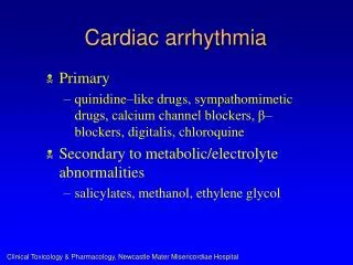
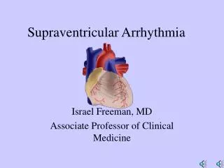
ARRHYTHMIA Cardiological Department Li Hongbo. Anatomy of the conducting system. ELECTROPHYSIOLOGIC PRINCIPLES. Automaticity:

E N D
ELECTROPHYSIOLOGIC PRINCIPLES • Automaticity: • the property of a cardiac cell has depolarization spontaneous during phase 4 of the action potential. normally observed in the sinus node; the specialized fibers of the His-Purkinje system; some specialized atrial fibers .
ELECTROPHYSIOLOGIC PRINCIPLES • Excitability • Refractoriness-the period of recovery that cells can be reexcited by a stimulus after being discharged. • Absolute refractory period • Effective refractory period • Relative refractory period
ELECTROPHYSIOLOGIC PRINCIPLES • Conductivity • the cardiac conducting system included sinus node; interatria; atrioventricular node; right and left bundle branch; purkinje system. • The conduction speed is fastest through purkinje system and is slowest through the atrioventricular node.
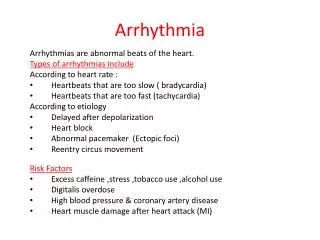
Classification of arrhythmia • Pathogenesis of arrhythmia • Disturbances of impulse formation • Sinus nodal disturbance • Ectopic rhythm • Disturbances of impulse conduction • physiological • pathological
Classification of arrhythmia • Heart rate of arrhythmia • Bradyarrhythmias • Tachyarrhythmias
Normal sinus rhythm • The normal sinus rhythm is defined as sinus rhythm with a heart rate between 60 and 100 beats/min. • The P wave is negative in lead aVR and positive in lead II
Sinus tachycardia • With a heart rate exceeding 100 beats/min , Generally is between 100 and 180 beats/min • Sinus tachycardia is an arrhythmia, but not necessarily an abnormal rhythm. • The following conditions can develop sinus tachycardia: Such as exercise, anxiety, fever, hyperthyroidism, anemia, myocarditis and some drugs.
SinusBradycardia • ECG characteristics: sinus rhythm is present and the heart rate is less than 60 beats/min • As a normal variant many normal and older people have sinus bradycardia • Sinus bradycardia commonly occurs in the following conditions: trained athletes, during sleep, Hypothyriodism, drugs or sick sinus syndrome • Most people with sinus bradycardia have no symptoms. If the patient has chronic sinus bradycardia cause symptoms, an electronic pacemaker may be needed.
Sinus Arrhythmia • The definition of sinus arrhythmia is sinus rhythm with an irregular rate. • The maximum sinus cycle length minus the minimum sinus cycle length exceeds 0.12 sec • The most common cause of sinus arrhythmia is respiration • This arrhythmia is a normal finding in children and teenagers.
Sinus Arrest or Sinus Pause • Sinus arrest or pause is recognized by a pause in the sinus rhythm. • The P-P interval of the pause does not equal a multiple of the basic P-P interval. • Sinus arrest or pause will lead to cardiac arrest with asystole unless the sinus node regains function or some other pacemaker (escape pacemaker) takes over.
Sinus Arrest or Sinus Pause • Sinus arrest can be caused by hypoxia, myocardial ischemia, hyperkalemia, digitalis toxicity, and some drugs. • In elderly people the sinus node may undergo degenerative changes and fail to function effectively.
Sick Sinus Syndrome(1) • Sick sinus syndrome is a term that is applied to a syndrome encompassing a number of sinus nodal abnormalities • SSS encompasses both disordered SA node automaticity and SA conduction. • With marked sinus bradycardia, sinus arrest, sinus exit block or junctional escape rhythms • Bradycardia-tachycardia syndrome
Sick Sinus Syndrome (2) • EKG Recognition: • Inappropriate sinus bradycardia; sinus arrest • Bradycardia -tachycardia syndrome (sinus bradyarrhythmia and nonsinus tachyarrhythmia). • AF or Afi with a slow ventricular rate response in the absences of drugs • Escape rhythm in the setting of persistent sinus arrest or exit block
The term “ectopy” , “ectopic pacemaker”, “ectopic beat” are used to describe non sinus beats. • Ectopic beats can be premature: premature atrial contractions (PACs) premature AV junctional contractions (PJCs) premature ventricular contractions (PVCs)
Some definition about the premature beats • Coupling interval: refers to the interval between the premature beat and the preceding normal beat. • Compensatory pause: Compensatory pause indicates that the interval between the normal QRS complexes immediately before and immediately after the premature beat. A fully compensatory pause is exactly twice the basic R-R interval.
Premature atrial contractions (PACs) • Premature beats arising from somewhere in either the left or the right atrium but not in the sinus node. • The atria are depolarized from an ectopic site. • The ventricular depolarization is generally not affected by PACs.
Premature atrial contractions (PACs) • PACs have the following major features: 1. The beat is premature 2. The PAC is often preceded by a visible P wave. Occasionally the P wave may be “buried” in the T wave of the preceding beat. This P wave has different shape and/or different PR interval from the P wave seen with the normal sinus beats. . 3. With noncompensatory pause. 4. The QRS complex is normal. Occasionally, PACs will result in aberrant ventricular conduction so the QRS is wider than normal.
Premature junctional contractions (PJCs) • Premature beats arising from AV junction • The direction of atrial depolarization is from bottom to top, just opposite to direction of normal sinus rhythm. • The P wave is upright in lead aVR and downward in lead II • QRS complex is normal. • Fully compensatory pause
P wave is upright in lead aVR and downward in lead II P wave will before, after, or buried in the QRS complex. P-R interval is less than 0.12’, R-P interval is less than 0.20’
Premature ventricular contractions (PVCs) • Premature beats arising in either the right or the left ventricle • The EKG characteristics of PVCs: • Aberrant in appearance. QRS complex is abnormally wide, usually 0.12 sec or more. T wave and the QRS complex usually point in opposite directions. 2. Have fully compensatory pause. 3. Interpolated PVC: A PVC falls between two normal beats
Ectopic Tachycardia • A run of three or more consecutive premature beats • paroxysmal atrial tachycardia • paroxysmal AV junctional tachycardia • Both of them are called supraventricular tachycardia 3. paroxysmal ventricular tachycardia
Paroxysmal supraventricular tachycardia (PSVT) • Including paroxysmal atrial and AV junctional tachycardia • Mechanism: • Reentrant • Automatic
Paroxysmal supraventricular tachycardia (PSVT) • EKG characteristics • The heart rate is between 150 and 240 beats/min • Extremely regular • P waves may or may not be visible. When seen, they are different from the P waves with normal sinus rhythm. • The QRS complexes are normal A wide QRS complex will be seen if the patient has an underlying bundle branch block or if the PSVT induces a “rate -related” bundle branch block.
Paroxysmal ventricular tachycardia (PVT) • A run of three or more consecutive PVCs • May occur as a single isolated burst, recurrent, or may persist for a long run
Paroxysmal ventricular tachycardia (PVT) • EKG characteristics • The heart rate is generally between 140-200 beats/min • The QRS complex is abnormally wide, usually 0.12 sec or more. • AV dissociation • Fusion beats, Sinus capture beats