
Interactive 3D-CT Organ Segmentation Tool using Principal Curve Projection
Develop an interactive tool for semi-automatic segmentation of organs in 3D-CT scans to enhance efficiency and accuracy in radiation therapy planning.

Interactive 3D-CT Organ Segmentation Tool using Principal Curve Projection
E N D
Presentation Transcript
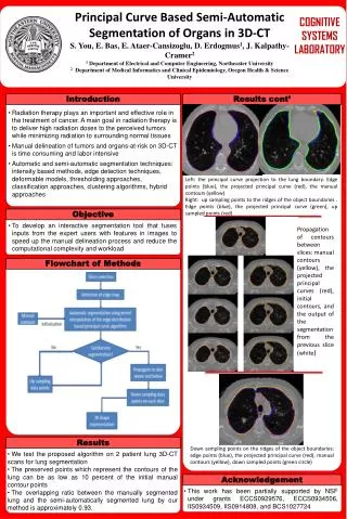
Principal Curve Based Semi-Automatic Segmentation of Organs in 3D-CT S. You, E. Bas, E. Ataer-Cansizoglu, D.Erdogmus1, J. Kalpathy-Cramer2 1 Department of Electrical and Computer Engineering, Northeaster University 2 Department of Medical Informatics and Clinical Epidemiology, Oregon Health & Science University COGNITIVE SYSTEMS LABORATORY Introduction Results cont’ Introduction Results cont’ • Radiation therapy plays an important and effective role in the treatment of cancer. A main goal in radiation therapy is to deliver high radiation doses to the perceived tumors while minimizing radiation to surrounding normal tissues • Manual delineation of tumors and organs-at-risk on 3D-CT is time consuming and labor intensive • Automatic and semi-automatic segmentation techniques: intensity based methods, edge detection techniques, deformable models, thresholding approaches, classification approaches, clustering algorithms, hybrid approaches Left: the principal curve projection to the lung boundary. Edge points (blue), the projected principal curve (red), the manual contours (yellow) Right: up sampling points to the ridges of the object boundaries . Edge points (blue), the projected principal curve (green), up sampled points (red) Objective • To develop an interactive segmentation tool that fuses inputs from the expert users with features in images to speed up the manual delineation process and reduce the computational complexity and workload Propagation of contours between slices: manual contours (yellow), the projected principal curves (red), initial contours, and the output of the segmentation from the previous slice (white) Flowchart of Methods Results • We test the proposed algorithm on 2 patient lung 3D-CT scans for lung segmentation • The preserved points which represent the contours of the lung can be as low as 10 percent of the initial manual contour points • The overlapping ratio between the manually segmented lung and the semi-automatically segmented lung by our method is approximately 0.93. Down sampling points on the ridges of the object boundaries: edge points (blue), the projected principal curve (red), manual contours (yellow), down sampled points (green circle) Acknowledgement • This work has been partially supported by NSF under grants ECCS0929576, ECCS0934506, IIS0934509, IIS0914808, and BCS1027724


