Download
1 / 35
380 likes | 499 Views
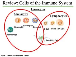
Santhakumar Manicassamy,1 Boris Reizis,2 Rajesh Ravindran,1 Helder Nakaya,1 Rosa Maria Salazar-Gonzalez,1,3 Yi-chong Wang,1 Bali Pulendran1,4*. Activation of β - Catenin in Dendritic Cells Regulates Immunity Versus Tolerance in the Intestine. Immune Cells. Tolerance vs. Immunity.

E N D
Santhakumar Manicassamy,1 Boris Reizis,2 Rajesh Ravindran,1 Helder Nakaya,1 Rosa Maria Salazar-Gonzalez,1,3 Yi-chong Wang,1 Bali Pulendran1,4* Activation of β-Cateninin Dendritic Cells Regulates Immunity Versus Tolerance in the Intestine
Pathogenesis of Inflammatory Bowel Disease In normal conditions, a balance is established between the generation of proinflammatory (TH1; TH2, TH17) and anti-inflammatory (TH3, Treg1) T cells Mediators indicated in blue (boldface) down-regulate inflammation, and those indicated in red promote inflammation.
Pathogenesis of Inflammatory Bowel Disease: Induction of Inflammatory Cascade Bacterial antigens inappropriately leak across the intestinal epithelial cell barrier. Tissue macrophages and dendritic cells (DCs) present these antigens to resident CD4+ T cells and activate proinflammatory T cells relative to regulatory T cells (Tr1, TH3, and NK-T cells) leading to excess proinflammatory cytokine release (IL-1, TNF-α) from the macrophages. Activation of the immune and vascular systems is regulated by nerve cells and their mediators (substance P). NK-T cell indicates natural killer T cell; TNF, tumor necrosis factor.
S1 Analysis of gene expression profiles using microarray analysis of purified intestinal lamina propria DCs (LP-DCs) and compared with those of splenic DCs (spl-DCs) mice. Several Wnt-ligands were up-regulated in LP-DCs as compared with spl-DCs. Consistent with this, reverse transcriptase polymerase chain reaction (RT-PCR) analysis showed that LP-DCs constitutively expressed several Wnt-ligands as compared with that of spl-DCs in the steady state (fig. S1)
Background • β-catenin, a central component in Wnt signaling, is widely expressed in hematopoietic stem cells, macrophages, DCs, and lymphocytes. • The Wnt–β-catenin pathway has been implicated in the differentiation of DCs from hematopoietic stem cells. • Activation of Wnt-Frizzled (fzd) receptor signaling causes translocation of β-catenin from the cytoplasm to the nucleus, where it interacts with T cell factor/ lymphoid enhancer factor (TCF/LEF) family members and regulates transcription of several target genes
S2 9. Mucosa10. Lamina propria11. Epithelium Assessment of whether the β-catenin pathway was active in intestinal Lamina Propria-DCs using the T Cell Factor/Lymphoid Enhancer Factor–lacZ reporter mice. In contrast to splenic-DCs, LP-DCs from TCF-reporter mice showed strong β-galactosidase expression, suggesting that β-catenin signaling pathway is constitutively active in intestinal DCs (fig. S2).
s3A To directly assess the role of β-catenin specifically in DC function, they crossed floxed β-catenin allele mice (β-catfl/fl) with transgenic mice (CD11c-cre) expressing the cre enzyme under the control of the CD11c promoter . This specifically abrogated β-catenin expression in DCs (fig. S3A).
Conditional targeting construct In this cartoon, the loxP sites are represented by arrowheads, the first and second exons by open boxes, introns by lines, and the targeted gene by the shaded box. Cre enzyme is introduced by transient transfection with a Cre-expressing plasmid CD11c under the control of CD11c promoter PCR is then used to find clones that are lacking the targeted gene but still have the first and second loxP sites, as illustrated by the next cartoon. The ES cells resulting from the partial recombination reaction illustrated above are said to have a floxed gene, meaning the gene of interest has one or more exons flanked by loxP sites (flox = flanked by loxP). When a mouse is made from these ES cells, it has an almost completely wildtype allele that can be knocked out by the expression of Cre inside its cells (assuming removal of the first exon results in a knockout).
S3B Distinct subsets of intestinal DCs and macrophages can be distinguished by the expression patterns of CD11c and CD11b (CD11c+CD11b– and CD11c+CD11b+ DCs and CD11c–CD11b+ macrophages) β-catenin was highly expressed and constitutively active in LP-DC subsets and LP macrophages in the small and large intestine (Fig. 1A and fig. S3B). In β-catDC−/− mice, β-catenin expression was abrogated in the CD11c+CD11b– and CD11c+CD11b+ DCs but only partially abrogated in CD11c–CD11b+ macrophages (fig S3B).
β-catenin signaling is constitutively active in intestinal APCs and regulates the induction of Treg cells and effector T cells. Figure 1A (A) Expression of β-galactosidase (representing β-catenin activity) by intestinal DCs and macrophages from SI-LP and LI-LP of TCF-reporter mice.
S4A Whether β-catenin signaling in DCs is critical for intestinal homeostasis. We compared the frequencies of Treg cells and TH17/TH1 cells in the intestine of β-catDC−/− and β-catfl/fl mice because intestinal DCs and macrophages play an important role in the induction of these subsets. β-catDC−/− mice had lower frequencies of Treg cells in the small intestine (SI)–LP, the large intestine (LI)–LP, and caecum, but not the spleen, as compared with that of β-catfl/fl mice (Fig. 1B and fig S4A).
Figure 1B (1B and 1C) Percentages of CD4+ T cells positive for FoxP3 (Treg cells), IL-17 (TH17), and IFN-g (TH1) isolated from Small Intestine-LP, Large Intestine-LP, Ceacum, and Spleen of β-catfl/fl and β-catDC−/− mice. Fluorescence activated cell sorting (FACS) plots Numbers in FACS plots represent % of cells positive for the indicated protein.
To determine whether the reduced frequencies of Treg cells observed in the LP of β-catDC−/− mice was due to increased frequencies of effector CD4 cells, we examined the frequencies of TH1 and TH17 in the intestine and periphery. S4B,C
Figure 1C They observed higher frequencies of TH17 and TH1 cells in the SI-LP and LI-LP but not in the spleen of β-catDC−/− mice as compared with that of the β-catfl/fl mice (Fig. 1C and fig. S4, B and C).
S5 β-catenin signaling in intestinal DCs is critical in maintaining the balance between Treg cells and CD4+ T effector populations CD4+ T cells isolated from SI-LP and LI-LP of β-catDC−/− mice showed elevated expression of the TH17 cell–associated mRNAs interleukin (IL)–17, IL-21, Rorgt, and the TH1 cell–associated mRNA interferon (IFN)–g (23, 24) as compared with the CD4+ T cells isolated from β-catfl/fl mice.
Figure 1D (D and E) Intracellular expression of FoxP3, IL-17, and IFN-g in naïve CD4+OT-II T cells stimulated to differentiate in vitro by intestinal APCs (CD11c+CD11b– and CD11c+CD11b+ DCs and CD11c–CD11b+ macrophages) isolated from β-catfl/fl or β-catDC−/− mice, in the presence of TGF-β (1 ng/ml
Fig. 2. β-catenin signaling in intestinal DCs promotes the expression of Raldh and suppresses the expression of proinflammatory cytokines. Figure 2A (A) Expression of Aldh1a1 and Aldh1a2 mRNA in CD11c+CD11b– and CD11c+CD11b+ DCs and CD11c–CD11b+ macrophages isolated from SI-LP of β-catfl/fl or β-catDC−/− mice. Retinal dehydrogenase (RALDH) catalyzes the oxidation of retinal to all-trans and 9-cis retinoic acid, which function as ligands controlling RAR and RXR nuclear receptor-signaling pathways.
S11 Because β-catDC−/− LP-DCs had reduced levels of vitamin A–metabolizing enzymes, we assessed whether addition of exogenous RA can rescue their defect in Treg induction. Addition of exogenous RA to β-catDC−/− LP-DCs did indeed enhance their capacity to induce Treg cells as well as β-catfl/fl DCs (fig S11). Thus, β-catenin– mediated signaling promotes Treg induction through the expression of vitaminA–metabolizing enzymes in DCs.
Figure 2B (B)Expression of Raldh protein by CD11c+CD11b– and CD11c+CD11b+ DCs and CD11c– CD11b+ macrophages isolated from SILP of b-catfl/fl or b-catDC−/− mice, as assessed by intracellular staining and flow cytometry.
LP-DCs had higher mRNA levels of the pro-inflammatory cytokines IL-23a and IL-6 and lower mRNA levels of the anti-inflammatory cytokines IL-10 and TGF-b, as compared with that of b-catfl/fl LP-DCs, from the SI and LI Figure 2C (C) Expression of Il-10, TGf-b1, Il-23a, and Il-6 mRNAs in CD11c+CD11b– and CD11c+CD11b+ DCs and CD11c–CD11b+ macrophages isolated from SI-LP of β-catfl/fl or β-catDC−/− mice.
β-catenin signaling in LP-DCs is critical for the induction of anti-inflammatory cytokines, which are crucial for the induction of Treg cells and suppression of TH1 and TH17 responses Figure 2D (D) Cytokine concentrations in the supernatants of CD11c+CD11b– and CD11c+CD11b+ DCs and CD11c–CD11b+ macrophages isolated from SI-LP of b-catfl/fl or b-catDC−/− mice after 24 hours. *P < 0.05; **P < 0.005.
Fig. 3. β-catenin activation in intestinal DCs is independent of commensals and induced by Wnt-signaling. Figure 3A (A and B) FACS plots representing the percentage of CD4+ T cells that express (A) FoxP3, IL-17, mice treated with or without Antibiotics
Figure 3B (B) IFN-g isolated from SI-LP of β-catfl/fl or β-catDC−/− mice treated with (Atx) or without (None) antibiotics.
Figure 3C (C) FACS plots showing the intracellular expression levels of β-galactosidase or β-catenin protein in CD11c+CD11b– and CD11c+CD11b+ DCs and CD11c–CD11b+ macrophages isolated from SI-LP of TCF-reporter mice treated with or without antibiotics.
Figure 3D & 3E (D and E) mRNA expression of (D) Fzd receptors and (E) Wnt ligands in CD11c+CD11b– and CD11c+CD11b+ DCs and CD11c–CD11b+ macrophages isolated from SI-LP of β-catfl/fl mice.
Figure 3F (F) mRNA expression levels of Wnt–β-catenin target genes Wisp1, Wisp2, and Axin1 in CD11c+CD11b– and CD11c+CD11b+ DCs and CD11c–CD11b+ Macrophages isolated from SI-LP of β-catfl/fl mice. Error bars indicate mean T SEM. Data are from one experiment representative of three.
Ablation of β-catenin expression in DCs enhanced inflammatory responses and disease in a mouse model of inflammatory bowel disease. Figure 4A β-catenin signaling in LP-DCs regulates intestinal homeostasis. A) Percent body weight of β-catfl/fl or β-catDC−/− mice treated with 2% DSS (Dextran Sulfate Sodium) for 7 days induced weight loss indicative of severe inflammation. Error bars indicate mean T SD.
Figure 4B (B) FACS plot representing percentage of CD4+ cells positive for IL-17 and IFN-γ isolated on day 8, from Ceacum and LI-LP of β-catfl/fl or β-catDC−/− mice treated with DSS for 6 days.
Figure 4C (C) Histopathological changes in colon tissue from β-catfl/fl or β-catDC−/− mice treated with or without 2% DSS treatment for 7 days. Areas of interest are infiltrations, edema (yellow arrows), goblet cells (black arrows), and basement membrane (green arrows).
In Summary • Demonstrated that a signaling pathway involving β-catenin in DCs regulates the balance between inflammatory versus regulatory responses in the intestine. • The present data demonstrate that β-catenin signaling in DCs is critical for maintaining DCs in tolerogenic state, via induction of various anti-inflammatory factors such as RA, IL10, and TGFβ. • Strategies that can activate β-catenin signaling specifically in DCs may be attractive means of controlling autoimmune diseases such as IBD via the induction of Treg cells and concomitant suppression of inflammatory effector T cells.