Download

1 / 69
690 likes | 859 Views
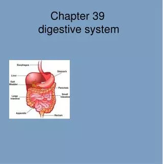
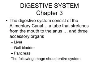
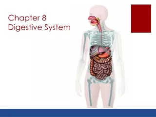
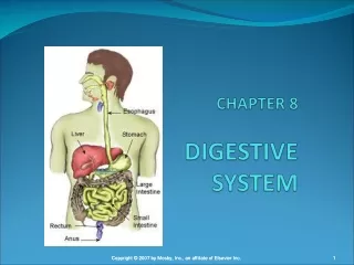
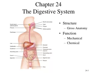
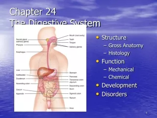
DIGESTIVE SYSTEM Chapter 3. The digestive system consist of the Alimentary Canal….a tube that stretches from the mouth to the anus … and three accessory organs Liver Gall bladder Pancreas The following image shows entire system. Alimentary Canal Structure.

E N D
DIGESTIVE SYSTEMChapter 3 • The digestive system consist of the Alimentary Canal….a tube that stretches from the mouth to the anus … and three accessory organs • Liver • Gall bladder • Pancreas The following image shows entire system
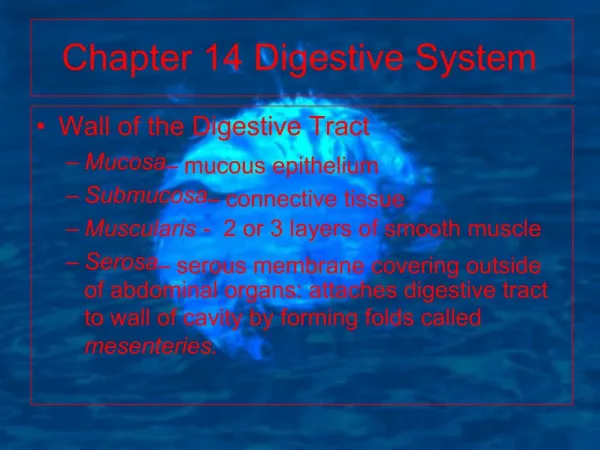
Alimentary Canal Structure • The Alimentary canal is organized into 4 functional layers: • Mucosa: • Submucosa • Muscularis • Serosa
Mucosa: • This is the site of mucous production • The mucous aids in transportation, protection and absorption of nutrients
Submucosa • It lies directly under the mucosa and contains VANL (vein, artery, nerve and lymphatic supply) • It is engaged in transportation of nutrients and cellular waste • It might also contain specialty glands
Muscularis • The muscularis is composed of two layers of smooth muscle • Circular layer • Longitudinal layer • These two layers works together rhythmically to create peristalsis to move food along.
Peristalsis • Peristalsis is the synchronous rhythmic contraction of the circular and longitudinal muscle • When circular contracts, the tube lengthens • When longitudinal contracts, the tube shortens
Serosa • The serosa is the most external layer • It secretes a viscous fluid that helps to reduce friction so that our parts can slip/slide past each other
From the Beginning • Let’s start from the beginning (oral cavity) and work our way to the finish (anus)
Structures for Study • Labia • Tongue • Teeth • Salivary glands • Palate A lot of structures are found in the mouth…these are the ones we are going to examine
Function(s) • The lips are a sensory organ that detect temperature….reduce the risk of burning • In terms of digestion, labia also help to keep food in the mouth
Tongue • Gustatory organ: taste • Needed for food placement • Needed for proper swallowing • Library.thinkquest.org
Taste sweet salty bitter sour
Taste cont’ • And for some reason power point would not let me add to the last slide • Our fifth type of taste is umame (savory) and that occurs over the entire tongue • Taste is regulated by the taste buds
Taste Buds • They are embedded within the bumps on the tongues surface (papillae) • They detect chemical concentrations • The tip of the tongue detects sweet/salty • Laterally it detects sour • The rear of the tongue is bitter • The entire tongue is umame (savory)
Swallowing • When swallowing, the tongue pushes food to the rear of the mouth and places pressure on the epiglottis..thereby closing the windpipe so that we do not choke • Diseases that effect muscles: Parkinsons, ALS, Muscular Dystrophy carry a higher rate of aspiration…..why?
Teeth (Dentition) • Our teeth allows us to be omnivores • We have teeth for cutting, tearing and grinding • This ability to be an omnivore is what has made us successful as a species • Teeth are used for Mastication (chewing)
Tooth Development • Primary Teeth (deciduous ) • This is our first set….they erupt from 4-6 months and begin to fall out at 6-7 yrs • We have 20 primary teeth • 4 central incisors • 4 lateral incisors • 4 cuspids • 4 primary molars • 4 secondary molars
Secondary Teeth (permanent) • These are our adult teeth • All of them should be in by 21 • 32 of these • 4 central incisors 4 lateral incisors • 4 cuspids 8 bicuspids (premolars) • 4 primary molars 4 secondary molars • 4 tertiary molars
Salivary Glands • Three groups • Parotid: Cheek area on top of Masseter muscle • Submandibular: found at the floor of the mouth • Sublingual: found at the floor of the mouth
Function(s) of Salivary Glands • Secrete saliva • Saliva is mixed with food to create a bolus • Within saliva we find two prominent enzymes: • Salivary amylase: dissolves carbohydrates • Lingual Lipase: dissolves triglycerides
Enzymes • In our bodies, enzymes are protein based chemicals that speed up chemical reaction rate and decrease activation energy. • Because they are protein based, the can be denatured (change shape) and that disrupts function
Denaturing • Enzymes can be denatured with changes in • pH: to much acid or base can change their shape • Temperature: Increases in temperature can destroy enzymes…e.g., high fever
Role of Enzymes • In the digestive system, enzymes are utilized to reduce macronutrients into a more absorbable micronutrient • Okay…..back to the oral cavity
Palate • This is the roof of the mouth • Divided into the hard and soft palate • Functions to separate the oral and nasal cavities • Cleft Palate is developmental dysfunction of the palate
Divisions • Nasopharynx: back of the nasal cavity • Oropharynx: back of the throat • Laryngopharynx: top of throat • Pharynx functions as common passageway for respiratory and digestive systems
Esophagus • Tube extending from laryngopharynx to the stomach • First instance of sphincters in digestive systems • The esophagus has: • Upper Esophageal Sphincter • Lower Esophageal Sphincter
Sphincters • Sphincters are ring like structures that close tubes to controls flow they prevent backflow of food
Stomach Cont’ • The stomach is a j-shaped organ designed to hold 1 liter of food and fluid • It is divided into 4 regions (see image): • Cardia • Fundus • Body • Pylorus
Sphincters of Stomach • Cardiac or Lower Esophageal Sphincter: prevents back flow of food from stomach to the esophagus • Pyloric Sphincter: controls gastric (stomach) emptying
Gastric Pits • The stomach is lined with gastric pits • These pits increase surface area and contain specialty cells
Gastric Pit Cells • Mucous cells: secrete huge amounts of protective mucous • Parietal cells: secrete hydrochloric acid (HCl) and intrinsic factor…what are the functions of these chemicals? • Chief cells: secrete pepsinogen and gastric lipase (function?) • g- cells: secrete Gastrin ( function?)
Function(s) of the Stomach • The stomach takes the bolus from the mouth and mixes it with gastric juice to create chyme • It allows absorbs some nutrients…water • Food typically stays in the stomach for 1-3 hours • Describe gastric bypass surgery
Divisions of the Small Intestines • Duodenum: first 15 or so inches of the S.I. it is the where the accessory organs enter the digestive system. It is the workhorse of the gut • Jejunum: middle third. Site of nutrient absorption • Ileum: the end of the S.I. get huge production of mucous in this division…and some absorption
Mesentary • The mesentary anchors the small intestines to the body. It holds a lot of VANL suppy for the digestive system