Principles of Sensory Coding
380 likes | 968 Views
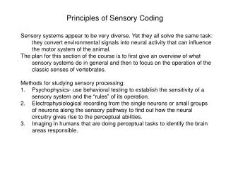
Sensory systems appear to be very diverse. Yet they all solve the same task: they convert environmental signals into neural activity that can influence the motor system of the animal.

Principles of Sensory Coding
E N D
Presentation Transcript
Sensory systems appear to be very diverse. Yet they all solve the same task: they convert environmental signals into neural activity that can influence the motor system of the animal. The plan for this section of the course is to first give an overview of what sensory systems do in general and then to focus on the operation of the classic senses of vertebrates. Methods for studying sensory processing: Psychophysics- use behavioral testing to establish the sensitivity of a sensory system and the “rules” of its operation. Electrophysiological recording from the single neurons or small groups of neurons along the sensory pathway to find out how the neural circuitry gives rise to the perceptual abilities. Imaging in humans that are doing perceptual tasks to identify the brain areas responsible. Principles of Sensory Coding
What do Sensory Systems Sense? There are two main functions of any sensory system: The detection of a signal. Weak signals can be detected without the animal being able to finely discriminate any of its features. Discrimination of some aspects of a sensory input. This is often referred to as estimation. What must be estimated from the input: 1. Qualitative features such as colour or odorant; this is often referred to as modality- what is it? 2. Quantitative features such as magnitude- often referred to as the intensity of a stimulus; 3. Temporal features such as duration or frequency of a signal; 4. Spatial location of a stimulus- where is it? Typically, all these aspects are estimated at once. A common strategy of sensory systems is to have separate neural pathways specialized for estimating different types of stimulus features. For example, the visual system analyzes colour, shape and movement in different brain regions.
How are these attributes represented in the brain? Modality: the most basic mechanism for identifying the nature of a sensory input is via labeled lines. What this means is that input from the optic nerve is always interpreted by the brain as visual input etc. This extends to much finer discriminations: the connections of “pain” and “touch” fibers in the somatosensory system are entirely different and electrical stimulation of either leads to the appropriate sensation. Intensity: the estimated intensity of a stimulus is not a linear function of the actual intensity. As shown in the graph below, the relation can be described as logarithmic or power law. The reason is intuitively easy to understand. Increases of a weak signal generate a larger perceived increase than increases of a strong signal- the percept saturates. A power law relation best describes the relation between stimulus strength and perceived stimulus intensity. Kandel, Schwartz and Jessel
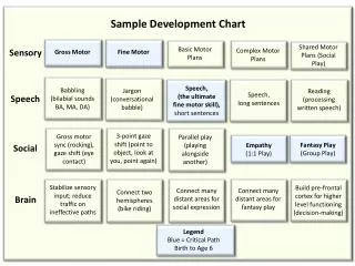
Temporal features: Onset time andDuration. The duration of a stimulus is estimated from the onset of the neural response and its duration. Typically there are neurons in sensory systems that only respond to the onset of a stimulus- these are generally referred to as phasic responders and they are good for estimating the time of occurrence of a signal. There are other neurons that respond throughout the stimulus presentation- tonic responders- these signal stimulus duration. Frequency. The frequency of a signal may be very important is some senses (audition). There are several ways to estimate this; one way is to make the neural response precisely time locked to the signal. Kandel et al. Bear et al.
Location. A common principle is used for estimating where a stimulus is located- topographic mapping. This means that points close together on the sensory surface are represented close together in the brain. In the somatosensory surface this is called “somatotopy”, in the visual system “retinotopy” and in the auditory system “tonotopy”. Skin regions that are close together are mapped to adjacent regions of the cerebral cortex. Bear et al.
Getting Sense Input into the CNS Sensory input comes in many flavours. Information in the CNS all comes in the same currency- action potentials or spikes. The reason that the CNS uses only one way to transmit information is simple: it allows integration of different types sensory input and the connection of sensory input to motor output- all the neurons dealing with these different systems use the same “language” of spikes. The problem becomes: how to translate the different kinds of sensory input into spikes. In all cases this is done by specialized receptor cells (or parts of cells) in a process called sensory transduction. The transduction process depends on the nature of the signal. Chemoreception. Mechanoreception. Vibration reception. Light. The initial transduction process causes the receptor cell to depolarize and this leads to spike initiation in sensory afferent fibers that then convey this information to the brain.
Sensory Transduction Stretch gated channels (in the nerve terminal of an afferent fiber) open and cause it to discharge. The fiber projects to the CNS. A chemical binds to the receptors on an afferent process, causes depolarization and discharge. A hair cell is activated by vibration, becomes depolarized and releases transmitter. The transmitter excites an afferent fiber; it discharges and this information reaches the CNS. Kandel et al. A photoreceptor is activated by light; its release of transmitter is reduced. After a series of complex interactions in the retina, ganglion cells discharge and send this information to the brain. The most important point is that the message sent by a receptor to the brain is in the form of a sequence of spikes- a spike train. What is this message?
Coding of sensory input All sensory input is represented in the firing patterns of populations of neurons. This is the most remarkable conclusion of sensory physiology. The representation is referred to as a population neural code. What is this code? The dominant theory since the Lord Adrian’s work in the early 20th century is that information is carried in a rate code- often a linear rate code. The intensity of a sensory stimulus is correlated with the firing rate (spikes/second) of a sensory afferent or neuron. This theory is well supported by data from some mechanoreceptors such as stretch receptors. In this experiment the firing rate of a neuron was recorded (over 1 second periods) while delivering stimuli with varying intensities. As you can see, the discharge rate varies in a linear fashion with intensity. This type of plot is often referred to as the tuning curve of a neuron. Kandel et al.
Other Possible Neural Codes 1 There are a number of problems with rate based codes. The most serious difficulty is that they are slow. Imagine a neuron discharging at 20 spikes/s in a fairly random (Poisson-like) manner (this is fairly typical for many cortical neurons). If it is activated it may increase its rate to 50 spikes/s; its target cells will have to wait for at least 1/2 second to figure out (reliably decode) that there has been a significant increase in firing rate. Our ability to detect novel input is far faster than this suggesting that other forms of coding must exist. I will list some candidate codes below in order of how firmly they are established. An important point is identifying a neural code requires more than merely showing that the information is present in a neurons discharge pattern. It must also be shown that the information is actually used by downstream neural circuitry- that it is decoded. In general this has proven to be very difficult to accomplish.
Other Possible Neural Codes 2 Coding with time: As you’ve already seen, neurons can become phase-locked to a stimulus. In this case, it is the time of occurrence of a spike that is the signal. This mechanism is well established in the auditory system. It is also possible that the time of occurrence of the first spike response to a stimulus carries most of the information. Coding with spike patterns: Many neurons (in thalamus, cortex and elsewhere) will, in response to sensory stimulation, produce bursts of spikes as well as trains of isolated spikes. It has been strongly argued, though not proven, that spike bursts are a separate coding mechanism. Coding with correlated activity in a neuronal population: It has been suggested that synchronized or correlated spiking activity is an important code. This type of code would permit a huge increase in coding capacity, but it has been difficult to conclusively demonstrate its occurrence.
Coding with Bursts of Spikes: Salient Sensory Input Spike bursts are brief, high frequency sequences of spikes. They are generated by different biophysical mechanisms including low threshold Ca2+ channels (thalamus) and interactions of a neuron’s soma and dendrites (pyramidal cells). Despite this, they may serve a common purpose- detection of important, brief and unexpected events. Here are two examples. In the retina reversing the direction of an object’s motion- an important and unexpected event, causes a synchronous spike burst of many retinal ganglion cells. In thalamus, neurons receiving input from the whisker afferents, burst when the rodent twitches its whiskers- a form of exploratory behaviour. Retina- motion reversal Thalamus- whisker twitching Schwartz, 2007 Fanselow, 2001
Coding with Bursts of Spikes: Reward Associated Signals Lin, 2008 Recordings from cells in basal forebrain; these cells project to cortex and might be related to learning. These cells respond to emotionally salient signals from any modality- emotionally salient means that the signals are associated with reward or punishment. When a neutral tone is presented the cells do not respond. What the figure shows is that, when the same tone has been paired with reward, it initiates a strong burst response (Hit)- but only when the rodent actually goes for the reward- there is no response to the tone if the animal does not go for the reward (Miss).
Receptive Fields I have been emphasizing neural coding as temporal patterns. Sensory physiologists must also take into account the spatial dimensions of neural coding. This is typically done by estimating a neuron’s receptive field. The RF is defined as that region of sensory space whose stimulation results in a change in discharge (usually firing rate) of the neuron Imagine that a patch of skin is represented at the left. A given receptor will be activated when the stimulus is presented in that region of skin where its terminal branches are found. This is that receptor’s RF. Many of these receptors will converge onto a neuron in the spinal cord or brainstem. This neuron will be excited when the skin is touched over a much larger region- it has a larger RF. Adding inhibitory interneurons into the circuit can reduce the size of the RF or complicate its response. Kandel et al.
For all sensory systems there is a common plan. Peripheral receptors respond to a specific stimulus and convert it (directly or indirectly) into a spike train. The afferent fibers end in lower brain regions where they are processed. Quite often there are many parallel pathways present. When rapid responses are required, the processed information might go directly to a motor system. However, for more detailed analysis, the information proceeds to higher brain levels for further processing. In mammals and birds, the sensory input reaches the forebrain where it somehow results in the perception of complex patterns. We will now look in more detail at the classic senses. The Overall Plan of Sensory Systems The ascending pathways of the somatosensory system. Kandel et al.
Parallel Pathways for General and Communication Signals A very general principle of sensory coding is that communication signals have their own separate channels. An animal might encounter very different environments depending on where it is born; for example, a city rat and a country rat will likely encounter very different odors. So sensory systems need to adapt themselves to the experiences of different animals. In contrast, communication signals evolve over evolutionary time and are highly conserved for each species. For example, many animals (including rats) use pheremones to communicate gender etc. These are fixed and so the olfactory system does not have to “learn” about different pheromones; their detection can be built in. This distinction is also evident in the auditory system: speech and song versus environmental sounds. It is also obvious in the visual system: expressions on faces versus trees etc. The obvious differences in the nature of these signals is carried forward in the nervous system and we’ll encounter this repeatedly in this course.
The Somatosensory System The somatosensory system is subdivided into 3 subsystems: Exteroreceptors. These are the familiar receptors found in the skin that mediate the sub-modalities of touch, pain and temperature. These types of sensory input can mediate both rapid responses (e.g. reflexes) and reach the cerebral cortex and induce perception. Proprioceptors. Proprioceptor afferents are found in muscles and at joints; they mediate the detection of muscle stretch and the degree of extension at a joint. For the most part ,this type of sensory input is involved in motor control rather than perception. The two major classes of proprioceptive input come from (a) Golgi tendon organs and (b) muscle spindles. The latter are highly specialized muscles that act to detect the amount of stretch of the muscle. We will not discuss these afferents any further in this section of the course. Interoreceptors. Interoreceptors are found in internal organs and convey signals that include distension of the stomach, carbon dioxide concentration in the blood etc. This type of sensory input is obviously of great importance but difficult to study experimentally (in the CNS). We will not discuss this submodality any further.
Exteroreceptors 1 The receptors that transduce light or sound are highly specialized cells; in contrast, exteroreceptors are merely nerve endings in the skin. The cells that give rise to these nerve endings are found in ganglia just external to the spinal cord (spinal ganglia) or brainstem (trigeminal ganglion). Ganglion cells are bipolar: one of its axons extends to the skin while the other enters the spinal cord. Each spinal ganglion innervates a strip of skin known as a dermatome. Bear et al.
Exteroreceptors 2 There are many forms of exteroreceptors. The most important distinction is between touch or mechano-receptors and pain and temperature receptors. The nerve endings of pain and temperature receptors are simple and the axons are either unmyelinated (C type) or lightly myelinated. In the case of touch fibers the nerve endings are usually associated with some specialized structures such as hairs. The axons of touch fibers are heavily myelinated allowing them to conduct action potentials rapidly. The response of touch fibers depends on their associated structures. Bear et al.
Response Properties of Fine Touch Fibers The structure of the accessory tissue of the nerve ending determines its response. High frequency Pacinian receptors are good for detecting texture. Ruffini endings estimate the duration of contact etc. Bear et al.
Central Projections of Exteroreceptors The central projections of somatosensory afferents allows this system to be divided into two functionally distinct streams: (a) a phylogenetically older spinothalamic system and (b) a more recently evolved (especially in mammals) lemniscal pathway. Kandel et al.
The Spinothalamic System The spinothalamic system is a phylogenetically old system that conveys sensory input related to pain, temperature and touch to the brainstem as well as cortex. Unmyelinated fibers (C type) carry pain input to the spinal cord. These afferents use Substance P (SP) as a co-transmitter with glutamate. When activated, these fibers release SP both peripherally and in the spinal cord. SP is very potent is activating spinal cord neurons that transmit pain information. There are also lightly myelinated fibers that do not have SP and convey transient pain signals. Bear et al.
The Spinothalamic System 2 Bear et al. Note that ST axons cross in the spinal cord before they ascend. So that pain and temperature are represented on the contralateral cortex. Spinothalamic fibers terminate densely within the spinal cord itself. This is important for reflex functions such as the withdrawal reflex. The ST fibers also end in the brainstem; again this is important for both low level motor control and for activating the reticular system- this causes heightened attention to potentially dangerous input. Finally, the ST axons reach the thalamus. They reach both non-specific thalamic regions that activate large parts of association cortex and the thalamic nuclei specifically involved with perception of touch.
The Dorsal Column, Medial Lemniscal Fine Touch System Bear et al. The dorsal column ML system contains only myelinated axons conveying fine touch information. The central axons give off collaterals in spinal cord with the main axon ascending via the dorsal columns to the medulla where they terminate in the dorsal column nuclei: n. gracils (trunk and legs) and n. cuneatus (mostly upper limb).
The Dorsal Column, Medial Lemniscal Fine Touch System 2 Ventroposterior nucleus of the thalamus. Note that lemnsical fibers cross in the medulla so that touch is represented in the contralateral cortex. Bear et al.
The Dorsal Column, Medial Lemniscal Fine Touch System 3 Touch for the face is conveyed by a functionally similar system. The afferent fibers come from the trigeminal nerve and enter in the pons. Pain and temperature fibers from the face then descend to the descending trigeminal nucleus. The fibers that carry touch information terminate in the principal trigeminal nucleus. From this point on, the touch afferents from trunk and face merge. Bear et al.
The Dorsal Column, Medial Lemniscal Fine Touch System 4 Representation of touch in cerebral cortex. Bear et al.
The Dorsal Column, Medial Lemniscal Fine Touch System 5 The columnar organization of cortex (Mountcastle). A column of S1 has neurons all responsive to the same type of stimulus from the same region of the body. This appears to be a major principle for the organization of at least sensory and motor cortices. Bear et al.
The Rodent Vibrissae System:A Model System for the Study of Active Touch Ahissar, 2008 Figure 3. Cortical representation of the five whiskers of interest (experiment R20). In this color scheme, white corresponds to no activity above the spontaneous level. Erchova, 2004 Rodents have prominent “whiskers” or vibrissae. These represent a dominant touch sense for these animals that they use to localize and identify objects (walls, insects, etc) in their environment even in total darkness. There are a constant number of vibrissae in every rat; they are heavily innervated by trigeminal afferents. Each whisker has muscles (motor trigeminus) at its base and it uses them to “whisk” – therefore this is an active sense in which the rodent actively generates movements that result in sensory stimulation. The whisker afferents reach cortex in a standard manner (via thalamus) and terminate in Layer 4. But each whisker input terminates in the center of a discrete ring of cells and the entire column of cells is devoted to that whisker- so this part of the rodent cortex is called “barrel cortex”. Because it is very convenient to stimulate whiskers and record from barrel cortex, the vibrissae system has become the ideal system to study somatosensory processing and the effects of peripheral damage (removing a whisker) on cortical processing.
Arabzadeh, 2005 The Vibrissae Sense can Encode Texture Sandpaper Coarse Fine Vertical Whisker Velocity Horizontal Peri-Stimulus Time Histogram PSTH Whisker Receptor Afferent Barrel Cortex Pyramidal Cell Peripheral and Cortical neuron respond with different spike patterns to different sandpaper textures. This is presumably the basis of a rodent’s (and ours) ability to discriminate texture.
Ahissar, 2008 The Vibrissae Sensory System uses many encoding Schemes The identity of the whisker activated encodes the vertical location of an object. The Horizontal location is encoded by timing of the spiking response of whisker afferent cells. The radial location is encoded by the firing rate of the spiking response. Horizontal Location Vertical Location The Response of Trigeminal neurons to Active Touch Differs from their Response to Passive Touch