Download
1 / 47
640 likes | 1.19k Views
Biomolecular NMR Spectroscopy Methods and applications to proteins Robert Kaptein. Novosibirsk, November 2012. C ontents. Introduction to Biomolecular NMR Multidimensional NMR for proteins Resonance assignments Observables & structure restraints 3D Structure determination by NMR

E N D
Biomolecular NMR SpectroscopyMethods and applications to proteinsRobert Kaptein Novosibirsk, November 2012
Contents • Introduction to Biomolecular NMR • Multidimensional NMR for proteins • Resonance assignments • Observables & structure restraints • 3D Structure determination by NMR • Protein Dynamics • Example: Lac repressor, structure, dynamics, DNA interactions
5 Pros & cons of NMR in structural biology • Pros... • no need for crystal: • no crystal packing artifacts, solution more native-like • study of dynamics: • picosecond to seconds time scales, conformational averaging, chemical reactions, folding... • easy study of protein-protein, protein-DNA, protein-ligand interactions • Cons... • NMR structure determination is a bit slow.... • Need isotope labeling (13C, 15N) • solution NMR works best for MW < 50 kDa
NMR & Structural biology DYNAMICS F-helices F-helices Apo-CAP CAP-cAMP Dynamic activation of an allosteric regulatory protein Tzeng S-R & Kalodimos CG Nature (2009)
NMR & Structural biology TRANSIENT COMPLEXES Visualization of the Encounter Ensemble of the Transient Electron Transfer Complex of Cytochrome c and Cytochrome c Peroxidase Bashir Q. et al JACS (2010)
NMR & Structural biology EXCITED STATES Structure and Dynamics of Pin1 During Catalysis by NMR Labeikovsky W. et al JMB (2007)
NMR & Structural biology MEMBRANE PROTEINS Mechanisms of Proton Conduction and Gating in Influenza M2 Proton Channels from Solid-State NMR Hu F. et al Science (2010)
NMR & Structural biology IN-CELL NMR High-resolution multidimensional NMR spectroscopy of proteins in human cells Inomata K. et al Nature (2009)
Nuclear spin (rad . T-1 . s-1)
The NMR sample • Isotope labeling • 15N for small proteins < 15 kDa • 15N & 13C for larger proteins, up to 30-40 kDa • 15N, 13C & 2H for large proteins > 40 kDa • selective labeling (e.g. only methyl groups) • Sample - recombinant expression in E.coli • pure, stable and high concentration • 500 µL of 0.5 mM solution: ~ 5 mg per sample • preferably low salt, low pH
Amino acids Amino acids are usually referred to with either a three-letter or a one-letter code:
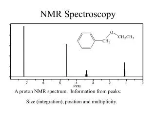
Protein 1H NMR spectrum Sensitivity: Signal to noise (S/N) Resolution: Line separation
Why multidimensional NMR • multidimensional NMR experiments • resolve overlapping signals • enables assignment of all signals • encode structural or dynamical information • enables structure determination • enables study of dynamics
2D direct dimension 3D indirect dimensions NxN FIDs of N points N FIDs of N points t2 t3 t1 mixing t1 t2 mixing mixing FID FID nD experiment 1D single FID of N points t1 FID
direct dimension indirect dimensions 1D 2D 3D mixing preparation preparation evolution evolution evolution N FIDs of N points t1 NxN FIDs of N points acquisition acquisition acquisition 1 FID of N points t2 t1 t1 t2 t3 mixing preparation mixing nD experiment
Encoding information mixing/magnetization transfer spin-spin interactions ???? proton A proton B
Encoding information: mixing • NOE (Nuclear Overhauser Effect ) • magnetic dipole interaction • through space • distance dependent (1/r6) • NOESY -> distance restraints • J-coupling interaction • through 1-4 bonds max. • chemical connectivities • TOCSY, COSY -> assignment • conformation dependent
COSY TOCSY J-coupling interaction transfer over several J-couplings, i.e. multiple steps over spin-coupled network t1 t2 t1 J-coupling interaction transfer over one J-coupling, i.e. max. 3-4 bonds t2 mlev FID FID Homonuclear 1H NMR NOESY magnetic dipole interaction crosspeak intensity ~1/r6 up to 5-6 Å tm t1 t2 FID
NOE effect |ββ> WS |βα> • Dipolar interaction • cross-relaxation • transient NOE build-up • proportional to 1/r6 NOESY W0 tm t1 t2 WI FID WI W2 |αβ> WS |αα>
(F1,F2) = ωA, ωB (F1,F2) = ωA, ωA A (ωA) A Cross-peak Diagonal B (ωB) B NOESY ~Å proton B t2 proton A t1 tm FID A (ωA) A ωA ωB F1 ωA F2
diagonal HN HN cross-peak NOESY (or 2D NOE) • Uses dipolar interaction (NOE) to transfer magnetization between protons • cross-peak intensity ~ 1/r6 • distances (r) < 5Å
COSY & TOCSY COSY o TOCSY *
Heteronuclear NMR • isotope labeling (expression in E.coli) • measure frequencies of different nuclei; e.g. 1H, 15N, 13C • no diagonal peaks • mixing only via J 1H 15N
Heteronuclear NMR HSQC (heteronuclear single quantum coherence) t2 J-mix block J-mix block 1H FID t1 15N DEC 1JNH 1JNH 15N (ω15N) 1H 1H (ω1H) (F1,F2) = ω15N, ω1H
x x y Δ Δ x x Heteronuclear magnetization transfer • INEPT • Insensitive Nuclei Enhanced Polarization Transfer • Enhancement factor: γH/γX (i.e. 10 for X = 15N) • Magnetization transfer through J-coupling • Chemical shift refocussed Δ = 1/(4JHN) 1H 15N
Iz α γB1 Product operator formalism z • Pulse (x) Izcosα + Iysinα • Chemical shift evolution Iycos(ωIt) + Ixsin(ωIt) • J-coupling evolution Iycos(πJISt) + 2IxSzsin(πJISt) y x Clockwise rotations !!! USEFUL RULES cos2α + sin2α = 1 cos2α - sin2α = cos2α 2cosαsinα = sin2α
Heteronuclear magnetization transfer Refocused INEPT y Δ Δ 1H Δ Δ 15N Δ = 1/(4JHN)
15N HSQC • Backbone NH • Side-chain NH and NH2
3D NMR • Double resonance • Two 1H frequency axes and one heteronuclear axis • 3D NOESY-15N-HSQC • 3D NOESY-13C-HSQC • 3D TOCSY-15N-HSQC • .... • Triple-resonance • Three different frequency axes (i.e.1H, 15N and 13C) • HNCA • HNCACB • HNCO • ....
3 HN atoms same δHN Same δHN different δ15N TOCSY HSQC F2 F3 1H → 1HN → 15N → 1HN t1 t2 t3 F1 FT 3D TOCSY-[1H-15N]-HSQC 1HN 1H 15N
3D TOCSY-[1H-15N]-HSQC strip 1H 1HN 15N [1H,15N]-HSQC projection
Triple resonance NMR HNCA t3 J-mix block J-mix block 1H FID J-mix block J-mix block t2 15N DEC t1 13C (aliphatic) 1JNCa(i) 1JNCa(i) 1JNH 1JNH 15N 1H 13C(ω13C) 15N (ω15N) 1H (ω1H) 2JNCa(i-1) 2JNCa(i-1) (F1,F2,F3)= (ω13Ca(i), ω15N(i), ω1H(i)) & (ω13Ca(i-1), ω15N(i), ω1H(i))
Sequential assignment • strips of 3D HNCA spectrum (15N dimension ⊥ to screen) 13Cα (i-1) 13Cα (i) 1HN (i)
Key concepts multidimensional NMR • resolve overlapping signals • mixing/magnetization transfer • NOESY, TOCSY, COSY • HSQC • 3D double resonance (3D NOESY-HSQC, 3D TOCSY-HSQC) • 3D triple resonance (e.g. HNCA)