Download
1 / 21
220 likes | 292 Views
What is Imaging and Radiation?. # radioactive#overplayed. The BASICS. Anatomic imagine began at the turn of the century with Roentgen's discover of x-rays in 1896 Advancements have allowed physicians (scientists) to look inside the body without the trauma and risk of explanatory surgery.

E N D
What is Imaging and Radiation? #radioactive#overplayed
The BASICS • Anatomic imagine began at the turn of the century with Roentgen's discover of x-rays in 1896 • Advancements have allowed physicians (scientists) to look inside the body without the trauma and risk of explanatory surgery.
Modern Techniques • Traditional x-rays • Computer Tomography (CT) • Magnetic Resonance Imaging (MRI) • Dynamic Spatial Reconstruction (DSR) • Digital Subtraction Angiography (DSA) • Positron Emission Tomography (PET)optical Coherent Tomography (OCT) • Echo-planar MIR/Ultrasound
Radiation Basics • Radiation is energy that comes from a source and travels through any kind of material and through space • Ionizing Radiation produces charged particles (Ions) • Ionizing radiation is produce by unstable atoms; unstable atoms are said to be Radioactive • To reach stability, these atoms give off energy; called Radiation
Types of Radiation • One typically encounters one of Three types: Alpha, Beta and Gamma Radiation • Neutron radiation is also encountered in nuclear plants, high altitude flights and emitted from industrial radioactive sources
Alpha Radiation • Heavy, very short range particle and actually an ejected helium nuclease • Characteristic; • Radiation Not able to penetrate human skin or clothing • Emitted materials can be harmful to humans • Very short distance; few inches
Beta Radiation • Light, short range particle, and ejected electron • Characteristics: • May travel several feet in air and moderately penetrating • Can penetrate human skin to germinal layer • May be harmful if deposited internally • Clothing provides some protection
Gamma Radiation • Or X-Rays, very long range, penetrating electromagnetic radiation • Characteristics: • Able to travel many feet in air and many inches in human tissue • Sealed radioactive sources that emit gamma radiation and an external hazard to humans • Visible light, radio waves and ultraviolet light • Electromagnetic radiations differ only in about of energy they have • Gamma/X-rays have most energetic • Clothing provides little shielding
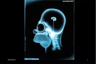
Radiography • Radiography, known by most as X-Ray, uses electromagnetic radiation to make images of your bones, teeth, and internal organs • X-rays allow DRs to take pictures inside the body • They Type of X-Ray depends on what part of the body needs examining and the purpose of the scan • Different tissues absorb different amounts of radiation • bones=dense, absorb X-Rays well; Appear White • Soft tissues; skin, fat, muscles and organs allow rays to pass, appear in varying shades of gray • Air appears black; lungs
Uses are as follows • Determine whether a bone is chipped, dislocated or broken (Fractured) • Evaluate Joint injuries and bone infections • Diagnose/monitor degenerative conditions; arthritis • Screen for lung and heart disease • Find and treat artery blockage • Diagnose cause of persistent couching or chest pain • Check from broken ribs/punctured lung • Evaluate unexplained abdominal pain • Help locate objects; maybe swallowed by a child • Detect scoliosis • Evaluate infection of the sinuses • Locate dental problems • Detection of Cancer
Preparations • In general: • Undress area of body needing examination; may wear gown • Remove jewelry, glasses, metal objects; Why? • May need to wear a lead apron; to shield sex organs, Why? • Medium Contrast; Barium and Iodine
Results • Image films usually developed or viewed on-screen within minutes • Radiologist typically views and interprets the results • Sends findings to doctor and than explain results • RISKS: • What are some risks? • Amount of radiation exposed to is extremely low—cancer?
Looking Ahead • X-Ray Technology has highly advanced from 1895 • X-ray Technology has specialized its imaging; such as mammography and Computerized Tomography Scanning (CT) • Fluoroscopy; real-time imaging of body on video • X-Ray Therapy: kill cancer cells and shrink tumors • Thoughts?
Typically the views of the body by these technologies are: • Transverse • Sagittal or • Frontal sections • Sagittal and Frontal are less difficult to understand; however most CT & MRI images are transverse • Which is why understanding cross-section anatomy is highly important
Computerized Tomography (CT) • Patient placed into a scanner and is exposed to a narrowly focused x-ray beam • Radiation not absorbed is called Remnant Radiation • Data is collected by a computer and converted into Pixels (these make a digital picture) • Image Reconstruction is when the computer combines the pixels to form an image (cross-Section) • Takes over 262.000 pixels to form a single CT Image
A Contrast Medium is a dense liquid that prevents the passage of remnant radiation and appears white in a CT image • This process is used to help with hollow organs (Blood vessels) to see contrast • New CT technology is able to produce an image so fast that physiological motions, respiratory or cardiac movements, do not blur the image • Where are the most commonly used? • Chest • Abdomen • Pelvis • Soft tissues of the neck • brain
Magnetic Resonance Imaging (MRI) • Patient is placed into a scanner that produces a very powerful magnetic field • Affects the nuclei of the atoms in patient's body to align • Once aligned a Radiofrequency Pulse is used to establish a second field • Data is collected by a computer by sensors to form pixels image reconstruction • Can produces transverse, sagittal or frontal sections • When pulse ends the hydrogen protons release an energy signal to realign with first magnetic field • Time required is called Relaxation Time • Two relaxation times: T1 (1st second image) and T2 (any time)
Appearance of Different Tissues in CT & MRI Images *Only if iodinated contrast media is present; otherwise appears gray