Download
1 / 28
290 likes | 317 Views
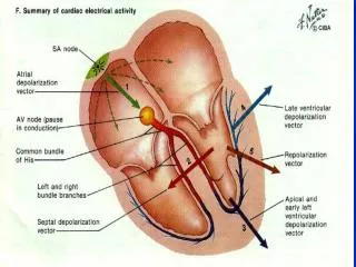
Normal EKG P wave: Atrial depolarization PR interval: < 0.20 sec QRS complex: ventricular depolarization QRS interval < 0.10 sec SA 0.10 – 0.12 supraventricular > 0.12 sec ventricular. Sinus Bradycardia. Sinus tachycardia. Pathophysiology: none. It’s a sign of something else.

E N D
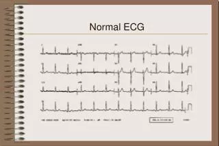
Normal EKG • P wave: Atrial depolarization • PR interval: < 0.20 sec • QRS complex: ventricular depolarization • QRS interval • < 0.10 sec SA • 0.10 – 0.12 supraventricular • > 0.12 sec ventricular
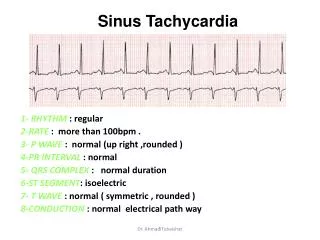
Sinus tachycardia • Pathophysiology: none. It’s a sign of something else. • Criteria: • Rate > 100 bpm • Rhythm: sinus • PR: < 0.20 • QRS: normal • Fever, exercise, hypovolemia, adrenergic stimulation.
Atrial Fibrillation/Flutter • Pathophysiology: Atrial impulses faster that SA. Reentry pathway • Criteria: • Rate: depends on atrial conduction • Rhythm: irregularly irregular • PR: cant diagnose. No P waves • QRS: narrow • CAD, CHF, vavular disease, hypoxia, drugs
PSVT • Pathophysiology: Reentry • Criteria: • Rate: > 150 bpm • Rhythm: regular • PR interval: cant see P waves • QRS: Narrow. • Accessory conduction pathway, CAD, COPD, CHF
Multifocal Atrial Tach • Pathophysiology: Increased areas of automaticity. • Criteria: • Rate: greater than 120 bpm • Rhythm: irregular • PR: variable (3 or more P waves) • QRS: narrow • COPD
1st degree block • Pathophysiology: slowed conduction • Criteria • Rate: slow < 60 bpm • Rhythm: regular • PR: > 0.20 second • QRS: narrow • Drugs (av nodal blockers, β-blockers, Ca+2 channel blockers, inferior MI.
2nd degree block I • Pathophysiology: AV node • Criteria • Rate: Regular or < 60 bpm • Rhythm: Regular with pause • PR: Progressive lengthing • QRS: narrow (until dropped) • Drugs, parasympathetic tone, RCA
2nd degree block II • Pathophysiology: Below the AV node • Criteria • Rate: Different between atrial and ventricular • Rhythm: Atrial – regular, vent – irregular • PR: normal • QRS: narrow (dropped beat) • ACS (LCA)
3rd degree block • Pathophysiology: no impulses from Atria • Criteria • Rate: Atrial – 60, ventricular – 40 • Rhythm: regular • PR: none • QRS: narrow or wide • ACS (LCA)