Download
1 / 49
490 likes | 873 Views
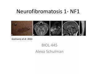
UNIVERSITÀ CATTOLICA DEL SACRO CUORE Facoltà di Medicina e Chirurgia “ A. Gemelli ” - ROMA ISTITUTO DI CLINICA PEDIATRICA Direttore: Prof. Riccardo Riccardi. CANCER SUSCEPTIBILITY AND MANAGEMENT IN CHILDREN WITH NEUROFIBROMATOSIS TYPE 1. Second primary cancer and familiar cancer syndromes.

E N D
UNIVERSITÀ CATTOLICA DEL SACRO CUOREFacoltà di Medicina e Chirurgia “A. Gemelli” - ROMAISTITUTO DI CLINICA PEDIATRICA Direttore: Prof. Riccardo Riccardi CANCER SUSCEPTIBILITY AND MANAGEMENT IN CHILDREN WITH NEUROFIBROMATOSIS TYPE 1 Second primary cancer and familiar cancer syndromes Rome, 27 January 2012
NF1 - EPIDEMIOLOGY • NF1 is one of the most common genetic disorders in the general population • Population prevalence: 1 of 3.500 live births • Sex ratio 1:1 • All racial groups are equally affected
NF1: A MULTISYSTEMIC GENETIC DISORDER • Autosomal dominant disorder • Many affected persons inherit the disorder, however 30 – 50% of new cases result from a spontaneous genetic mutation of unknown cause • Variable phenotypic expression: signs can range from benign cutaneous manifestations to life-threatening or sever disfigurement • Complete clinical manifestationsat 8-10 years of age • Symptoms and severity of the disorder may vary among members of an affected family
NF1: A TUMOUR-SUPPRESSOR GENE NF1: tumour-suppressor gene located on chromosome 17q11.2 NF1 encodes neurofibromin, a cytoplasmic protein that is expressed in neurons, schwann cells, oligodendrocytes, astrocytes and leukocytes Neurofibromin is a negative regulator of the Ras oncogene, the inactivation of which leads to cell proliferation and tumour development
NF1- DIAGNOSTIC CRITERIA Two or more of the features listed: • Six or more café au lait macules • larger than 5 mm in greatest diameter in prepubertal individuals • larger than 15 mm in greatest diameter in postpubertal individuals • Freckling in the axillary or inguinal regions • Optic pathway glioma • Two or more Lisch nodules • A distinctive osseous lesion, such as sphenoid dysplasia or thinning of the long bone cortex, with or without pseudoarthrosis • A first-degree relative with NF-1 according to the above criteria • Two or more neurofibromas of any type or 1 plexiform neurofibroma
NEUROFIBROMAS • Most common benign tumor of NF-1 • It can form at any place along a nerve • Three subtypes of neurofibroma : cutaneous, subcutaneous, and plexiform • Cutaneous lesions and subcutaneous lesions are circumscribed. These nodules may be brown, pink, or skin colored. They may be soft or firm to the touch • Plexiform neurofibromas are non circumscribed, thick, and irregular, and they can cause disfigurement by entwining important supportive structures cutaneous neurofibromas Plexiform neurofibroma
NF1 - BONE ABNORMALITIES • Scoliosis: 10-30% • Pseudarthrosis: 1 à 3 %
NF1 - NEUROPSYCHOLOGICAL DISORDERS • Mental retardation : 4 - 8 % • Learning disabilities: 40-60% • Epilepsy: 11 % • Headache
OTHER CLINICAL FEATURES OF NF-1 • congenital heart defects • Hypertension. In NF1 it can also result from renovascular disease, tumors that secrete vasoactive compounds, and coarctation of the aorta • Hypoglycemia • Fibromas in Iris • Glaucoma – rare
HEALTH SUPERVISION GUIDELINES FOR CHILDREN WITH NF1 * Biannual vision screening since the age of 7 years
NF1 AND NEOPLASTIC RISK • Patients with NF-1 have about a 3-15% additional risk of malignant disease in their lifetime • In NF1-related tumours, biallelic NF1 inactivation results in complete loss of functional neurofibromin activity. Most tumour suppressor genes are inactivated according to the two-hit model • Malignant tumours can arise in the nervous and non-nervous system in either childhood or adulthood • Malignanttumours are the most common cause of death in individuals with NF1 • The systematicmedical follow-up of children with NF1 isnecessary due to developments of malignanciesrelated to the disease Brems et al. Lancet Oncol 2009; 10:508–515
OPG IN CHILDREN WITH NF1 • Incidence of OPG in patients with NF1: 15 - 20 % • OPG are associatedwith NF1 in 50 %-70 % of cases • Diagnosisbelow the age of 6 years: 60-70 % of cases • Histologically, OPG are almost uniformly pilocytic astrocytoma (WHO grade I) • The majority of OPGs are slow growing • The prognosis is reportedly much better in children with NF1 than in those without (3-years progression free survival: 82% vs 42%) • Spontaneousregressionhasbeenreported in OPG associated with NF1
DIFFERENT FEATURES OF OPG IN CHILDREN WITH AND WITHOUT NF1 CHILDREN WITH NF1: - the most common site of involvement is the orbital nerve - bilateral optic nerve involvement is seen almost exclusively in NF1 patients - the tumor is smaller than in the non-NF patients and the original shape of the optic pathway is preserved - cystic components are uncommon CHILDREN WITHOUT NF1: - the chiasm and hypothalamus are the most common sites of involvement - cystic components are frequently seen Kornreich et al. Am J Neuroradiol 2001; 22:1963–1969
A SPECIFIC RADIOLOGICAL FEATURE OF NF1: UNIDENTIFIED BRIGHT OBJECTS (UBOs) • UBOSs: areas of increased signal intensity on T2-weighted images • 60% of affected children • lesions well circumscribed and nonenhancing, located in the brainstem, cerebellum, basal ganglia, and thalamus • the lesions are not space occupying and tend to disappear in adulthood
VISION SCREENING IN CHILDREN WITH NF1 • Ophthalmic screening is recommended in children with NF1 • The protocol includes visual acuity testing and fundoscopy every year + colour vision and visual fields when the child can co-operate • The first 6 years of life constitute the time of maximal risk for OPG development. • In children younger than 1 year • visual examinations may be unreliable! ASYMPTOMATIC CHILDREN < 8 YEARS: annual screening ASYMPTOMATIC CHILDREN > 8 YEARS: screening every 2 years until 18 years of age Listernick et al. Ann Neurol 2007;61:189–198
MRI: USEFUL SCREENING TOOL IN CHILDREN WITH NF1? • Some specialists recommend an initial MRI study to determine if an optic glioma is present when the diagnosis of NF1 is made, based on the difficulty in detecting visual changes in young children • Although potentially helpful in establishing an early diagnosis,based on the benign nature of most OPG and the need to sedate the child neuroimaging of the brain may not be warranted • Neuroimaging of asymptomatic children has not been shown to improve clinical • outcomes nor does early detection reduce the incidence of visual loss due to OPG • - MRI does not predict clinical behaviour or preclude the later development of an OPG • - Incidentally identified NF1-associated OPGs rarely require treatment
AN INTEGRATED APPROACH TO THE TREATMENT OF OPGs • Do we need to treat? • If yes, when? • And how?
OPG IN CHILDREN WITH NF1 - MANAGEMENT • Absolutely mandatory pre-therapeutic multidisciplinary discussion • Let’s be careful about the real need for treatment • Imaging is not the only criteria to start the treatment • We treat a child not an MRI • Treatment in patients with clinical and/or radiological progression (visual progression, defined as a two-line decrement in visual acuity, is an indication for chemotherapy) • Surgery is reserved for large tumors causing mass effect or hydrocephalus and tumors confined to the orbit or unilateral optic nerve • When a non surgical treatment is needed, decision should take into account: • Age • NF1 • Tumour site • Tumour volume
etoposide increased risk of second tumours !!!
RADIOTHERAPY IN CHILDREN WITH OPG AND NF1 • In chidren with NF1 RT is not recommended leading to serious complications: • Increased risk of second tumors • Neurovascular sequelae, moyamoya syndrome
SECOND CANCERAS IN IRRADIATED AND NON IRRADIATED NF1 PATIENTS Sharif S et al. J Clin Oncol. 2006 1;24:2570-5.
TARGETED THERAPY • Targeted therapy is addressed to key pathways involved • with the development of tumors in NF1: • Ras/mitogen-activated protein kinase (MAPK) • Akt/mammalian target of rapamycin (mTOR)
Recently, mTOR inhibitors such as everolimus have shown encouraging benefit for patients with subependymal giant cell astrocytoma
MALIGNANT PERIPHERAL NERVE SHEATH TUMOUR IN CHILDREN WITH NF1
MPNST IN CHILDREN WITH NF1 • The lifetime risk of a malignant peripheral nerve sheath tumour (MPNST) in patients with NF1 is 8–13% • Annual incidence: 0.16% (compared with 0.001% in the general population) • In patients with NF1 MPNSTs are difficult to diagnose because many of these patients have multiple benign tumors of the peripheral nervous system • Most NF1-associated MPNSTs arise within preexisting plexiform neurofibromas and may not produce symptoms until distant metastases occurr Plexiformneurofibromas that become painful or begin to grow rapidly should be investigated for the risk of malignant transformation!!! Cecen et al, Childs Nerv Syst (2011) 27:1885–1893
MPNST show increased glucose uptake on FDG-PET FDG-PET can differentiate most benign neurofibromas from MPNSTs Brems et al. Lancet Oncol 2009; 10:508-515
MPNST - MECHANISMS OF ONCOGENESIS • Inactivation of both NF1 alleles is thought to be sufficient to form a benign neurofibroma but not an MPNST • Most MPNSTs have a complex karyotype with many gains and losses of chromosomal material • The tumour suppressor genes CDKN2A and TP53 have an important role in the malignant transformation of neurofi broma to MPNST Brems et al. Lancet Oncol 2009; 10:508-515
MPNST IN NF1 PATIENTS: TREATMENT AND PROGNOSIS • Complete surgical resection is the mainstay of the successful treatment of MPNSTs • Radiotherapy can improve local control • Poor prognosis in children with NF1 compared to sporadic cases (5-year survival rate 21% vs 42%): • occurrence of larger, more invasive, and less resectable tumors in NF1 patients • tumours less responsive to chemotherapy (17% vs 55% in non-NF1 cases) • potential biological differences between MPNST patients with and without NF1
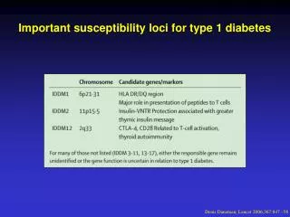
Embryonal rhabdomyosarcoma (bothyroid type) protruding from the vaginal meatus RMS IN CHILDREN WITH NF1 • The prevalence of RMS associated with NF1 is about 20 times higher than that in the general population • The pathogenesis of RMS in NF1 patients appears less clear than for MPNST • RMS arising in NF1 patients tend to have peculiar features: • embryonal histotypes • genitourinary tract or pelvic (paravesical) location • onset in early age
CONCLUSION • The development of malignanciesis a feature of NF1 • The clinicalcourse of malignancy in NF1 isoftendifferent from that of similartumortypes in the general population • Careful follow-up in patients with NF1 isrequired to enable the earlydiagnosis of malignancies
CASE REPORT 20 days-old child is brought to the pediatrician for a check up Clinical examination: healthy baby 7 café au lait macules larger than 5 mm in greatest diameter Family history: father and paternal grandmother with NF1 IS THE CHILD AFFECTED BY NF1?
NF1- DIAGNOSTIC CRITERIA Two or more of the features listed: • Six or more café au lait macules • larger than 5 mm in greatest diameter in prepubertal individuals • larger than 15 mm in greatest diameter in postpubertal individuals • Two or more neurofibromas of any type or 1 plexiform neurofibroma • Freckling in the axillary or inguinal regions • Optic pathway glioma • Two or more Lisch nodules • A distinctive osseous lesion, such as sphenoid dysplasia or thinning of the long bone cortex, with or without pseudoarthrosis • A first-degree relative with NF-1 according to the above criteria
HEALTH SUPERVISION GUIDELINES FOR CHILDREN WITH NF1 * Biannual vision screening since the age of 7 years
CLINICAL HISTORY • At 4 years of age: occurrence of headache and reduction of visual acuity • Brain scan showed a optic pathway mass, confirmed by IRM BIOPSY? The association between OPG and NF1 is a well-knownphenomenon. Diagnosis of OPG based on IRM.
TREATMENT CLINICAL AND RADIOLOGICAL EVOLUTION • Treatment according to SIOP LGG 2004 protocol (start on 28/05/2005; end on 18/06/2006) • Stable disease until July 2007 • MRI on 25/09/2007: • increase of the cystic component, without mass effect • no progressive visual impairment • MRI on 07/01/08: • reduction of the cystic component • no progressive visual impairment TREATMENT ? WAIT AND SEE
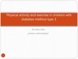
EXAMPLES OF SPONTANEOUS REGRESSION - 1 Sagittal MRI T2 flair scan of brain - at six months of age showing huge mass in optic chiasmatic region (a) - at four years of age showing significant regression of optic chiasmatic mass (b)
EXAMPLES OF SPONTANEOUS REGRESSION - 2 Coronal (a) and axial (b) T1 weighted MRI imaging after contrast administration, disclosing a chiasmatic tumor, performed when the boy was 2 years old. Coronal (c) and axial (d) T1 weighted MRI imaging after Gadolinium administration, 9 months later,demonstrate the complete regression of the chiasmatic lesion.
LET’S BE CAREFUL ABOUT THE REAL NEED FOR TREATMENT! • Imaging is not the only criteria to start the treatment ! We treat a child not an IRM • Treatment in patients with clinical and/or radiological progression
OUTCOME To date, no specific therapy is available for optic glioma – induced visual loss NEUROPROTECTION ? Now the child is 11 years old • off - therapy for 5.5 years • stable disease • visual acuity deficit persists
NERVE GROWTH FACTOR (NGF) • NGF is the first discovered neurotrophininvolved in the development and survival of sympathetic, sensory, and forebrain cholinergic neurons • given its actions favoring neuronal survival, NGF has been proposed for the treatment of some traumatic, ischemic, and neurodegenerative braindiseases • exogenousNGF has a neuroprotective effects on the visual system due to the presence of NGF receptors on the conjunctiva, cornea, as well as in the retinal pigment epithelium, photoreceptors, and retinal ganglion cells • NGF promotes: • protective activity against neural apoptosis • neuronal repair and • axonal regeneration
TREATMENT BY NGFpurified and lyophilizedfrom male mouse submandibular glands 1 mg of NGF diluted in 5 mL of saline solution was administered onto the conjunctiva of both eyes for 10 consecutive days 3 times a day
Five patients with OPGs and advanced optic nerve atrophy were assessed before and after a single 10-day course of 1 mg murine NGF topical administration by clinical evaluation, visual-evoked potentials (VEPs), and MRI After NGF treatment: - median VEPs amplitude showed a progressive increase from the baseline values (p < 0.01) - perception of spontaneous visual phosphenes was noted
Eye drop NGF administration appears to be a promising rescuing strategy for the treatment of children with optic glioma – induced visual loss NGF: A PROMISING RESCUING STRATEGY RISK TUMOUR PROGRESSION ? No significant change in tumor volume after NGF treatment was found
MRI of the brain • at baseline (A and B) • 4 months after NGF administration • (C and D) • No significant change in tumor volume • after NGF treatment