Download
1 / 51
520 likes | 729 Views
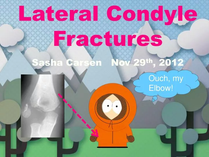
Lateral Condyle Fractures Sasha Carsen Nov 29 th , 2012. Ouch, my Elbow!. Agenda. Q ’ s Demographics, Mechanism Clinical Presentation Imaging Classification Treatment Complications Summary Q ’ s & A ’ s. 1.

E N D
Lateral Condyle Fractures Sasha Carsen Nov 29th, 2012 Ouch, my Elbow!
Agenda • Q’s • Demographics, Mechanism • Clinical Presentation • Imaging • Classification • Treatment • Complications • Summary • Q’s & A’s
1 A 36-year-old male presents for evaluation of left hand weakness. A current clnical photograph of his hand is shown in Figure A. His medical history is significant for the elbow injury shown in Figure B, which was treated non-operatively twenty-eight years previously. Current radiographic evaluation of the patients elbow will most likely reveal what deformity? 1. Cubitus valgus 2. Avascular necrosis of the lateral fragment 3. Fishtail deformity of the distal humerus 4. Fracture nonunion and a normal carrying angle 5. Myositis ossificans
2 Nonunion following a pediatric lateral condyle fracture has been associated with which of the following? 1. Ulnar nerve palsy 2. Radial nerve palsy 3. Heterotopic ossification 4. Parsonage Turner syndrome 5. Cubitusvarus
13 An 8-year-old boy falls on his right upper extremity and presents to the emergency room with the radiographs shown in Figures A and B. He has exquisite tenderness to palpation along the lateral aspect of his elbow. What additional radiographic view will likely demonstrate the maximum degree of fracture displacement? 1. External oblique radiograph 2. Internal oblique radiograph 3. Anteroposterior in maximum flexion 4. Anteroposterior in maximum extension 5. Lateral in maximum extension
4 A 7-year-old girl undergoes open reduction internal fixation of a displaced humeral lateral condyle fracture. Dissection around which portion of the fracture fragment should be avoided to protect its blood supply? 1. medial 2. lateral 3. superior 4. anterior 5. posterior
5 Figure A shows the radiograph of a 6-year-old girl after a fall on the playground. What is the most appropriate course of action? 1. Observation with treatment in a sling 2. Closed reduction and long arm casting 3. Closed reduction percutaneous pinning with k-wires 4. Open reduction internal fixation with k-wires 5. Open reduction with plate fixation
Lateral Condyle Fractures • Second most common # around the Elbow in children • Estimates range 12-20% of # Elbow • Average age 5-7 yrs • (Range roughly 4-10)
Mechanism • FOOSH • Elbow Extended & Supinated • Varus force, avulsion of Lateral Condyle • Flexed/Neutral/Pronated • Element of axial load directly transmitted through Radial head to capitellum • Likely some element of combined mechanism
Clinical Presentation & Eval • Trauma • Beware abuse if story questionable, red flags • Varus injury • Elbow pain, unable/unwilling to ROM or be examined • Tender Laterally, swelling, some ecchymosis • Neurovascular Exam • Acute not high risk like SCH
Imaging • Radiographs of affected Elbow • AP: Typically metaphyseal flake • Fragment typically postero-lateral • Internal Oblique view • Consider radiographs of contralateral elbow if questionable
US • In stable-appearing fractures there may be a role for US to define stability • Requires high level MSK-US experience/technique Sonographic differentiation of stable and unstable lateral condyle fractures of the humerus in children. Vocke-Hell AK, Schmid A Department of Paediatric Orthopaedics, Children's University Hospital, Basel, SwitzerlandJournal of Pediatric Orthopaedics. Part B [2001, 10(2):138-141]
Arthrography • Assess true articular displacement/reduction • More useful intra-operatively for attempted CRPP vs. ORIF/ORPP
CT & MRI • Both can add useful information, but significant downsides • CT • Does not visualize articular & non-ossified • ? Added benefit acutely • MRI • Excellent visualization • Cost, availability, SEDATION, time • How much will either change your plan?
Classification • Milch • Position of articular exit • Jakob • Stages of displacement Milch I Milch II Jakob 1 2 3
Jakob Classification • Type 1: Non-displaced fracture. Fracture line does not cross through the articular surface • Type 2: Minimally displaced. Fracture extends to the articular surface, but the capitellum is not rotated or significantly displaced • Type 3: Completely displaced. Fracture extends to the articular surface, and the capitellum is rotated and significantly displaced
Song et al., Closed Reduction and Internal Fixation of Displaced Unstable Lateral Condylar Fractures of the Humerus in Children. JBJS(Am). 90A9(12)2673.
Treatment Options • Non-Operative • LAC (may be LASplint first week) • Must be truly non- or minimally displaced • Must be followed weekly for displacement, and converted to operative Tx should displacement occur or signs of mal/non-union • Any concerns for F/U, should be considered relative indication for CRPP
Non-Operative Tx • 95 children undisplaced, or min displaced (<2mm), Tx w LAC/S. • Radiogrpahs - 3 views • 98% union by 7/52 • 2 pt’s req’d OR Bast, SC, Hoffer, MM, Aval, S. Nonoperative Treatment for Minimally and Nondisplaced Lateral Humeral Condyle Fractures in Children. Journal of Pediatric Orthopaedics. July/August 1998 - Volume 18 - Issue 4 - pp 448-450
Treatment Options • CRPP • Option for non-displaced who cannot be followed closely • Must be reduced anatomically • Best to assess with arthrogram • Mintzer et al., good results with CRPP for min displaced • Song et al., promising good results even with significant initial displacement, though required anatomic reduction
Treatment Options • Open RPP • Gold Standard • Anatomic reduction • Provisional fixation w clamp, etc. • Directly visualize the articular surface & Articular reduction • Stable Fixation
Surgery • Displaced fragment is typically posterolateral • Reduce (Closed vs Open) • Must be satisfied that articular surface is reduced (Direct viz vs. Arthrogramvs Scope)
Surgical Approach & Dissection • Direct Lateral • Kocher Interval vs. through split • Stay ANT. • Blood supply post. • Viz Articular surface
Fixation • 2 to 3 K-wires • Smooth & Threaded described • Preferably percutaneous
Fixation • Orientation of K-Wires dependent on # • K-wire transversely across capitellum/trochlea can prevent rotation • Bi-cortical
Post-op • LA Splint/Cast • Variable timeline, but begin ROM around 4/52 • Traditionally pins out 3-6/52 Three Weeks of KirschnerWire Fixation for Displaced Lateral Condylar Fractures of the Humerus in Children. TD. Phillip, Howard, Andrew W, Cole, William G., Hedden, Douglas M. Journal of Pediatric Orthopaedics: September/October 2001 - Volume 21 - Issue 5 - pp 565-569 • Watch for union • Warn of Lat column overgrowth, CubitusVarus & Tardy Ulnar n. Palsy
Treatment:Jakob Class Type 1: • Oblique radiographs may be necessary to confirm that this is not displaced. • LA Cast in 90deg • Follow very closely, watch for displacement
Treatment Type 2: • Displaced more than 2 mm on any radiograph (AP / Lateral / Oblique views)Reuquires Reduction • CRPP possible, but articular reduction must be anatomic. • If displaced, and the articular surface is not congruous, ORIF
Jakob Classification Type 3: • ORIF • (ORPP)
Complications & Concerns • Wide scar • Lateral subperiosteal new bone • Overgrowth lateral condyle • Pseudo-cubitus varus • Cubitus varus • Infection – rare
Growth Disturbances • Low incidence of growth arrest • Significant percentage develop Cubitus Varus • More common in non-op • Overgrowth of fractured region thought to be underlying etiology
Tardy Ulnar Nerve Palsy • Develops typically in 20’s • Ulnar n. Symptoms • Progressive weakness/atrophy • Some decr sensation • Typically resolves with decompression and transposition
Summary – Points & Pearls • Don’t miss the Dx – 3 views • Stable reduction/position – Maintain • Watch closely if no-op • Can take longer than SCH to heal (pins 3-6/52) • Be aware of complications – Early & Late • Mal-union can operate acutely until 8/52 • Non-union principles, compression w/ screw/BG • Tardy ulnar n. palsy – in 20’s
1 A 36-year-old male presents for evaluation of left hand weakness. A current clnical photograph of his hand is shown in Figure A. His medical history is significant for the elbow injury shown in Figure B, which was treated non-operatively twenty-eight years previously. Current radiographic evaluation of the patients elbow will most likely reveal what deformity? 1. Cubitus valgus 2. Avascular necrosis of the lateral fragment 3. Fishtail deformity of the distal humerus 4. Fracture nonunion and a normal carrying angle 5. Myositis ossificans
Figure B represents an acute lateral condyle fracture. Cubitus valgus deformity may occur due to a nonunion, malunion, or premature physeal closure. Acute neurologic injuries are rare with these injuries, however tardy ulnar nerve palsy (as demonstrated by the claw-hand deformity in Figure A) occurs late in the treatment and follow-up of lateral condyle fractures and usually is due to cubitus valgus. In addition to claw-hand deformity, other classical examination findings consistent with ulnar nerve palsy include Froment sign (compensatory thump IPJ flexion due to weak adductor pollicis) and Wartneberg sign (persistent abduction and extension of the small digit during active adduction due to weak interosseous and lumbrical musculature). Interosseous and/or first web space atrophy is another common finding. Storm et al reviewed elbow deformities after fracture. With regards to lateral condyle fractures, they state that the most common sequela in the setting of nonunion with displacement is the development of progressive cubitus valgus deformity. Valgus deformity can place an individual at risk for the developement of tardy unlar nerve palsy. This is a well-known late complication, occuring on average 22 years after initial injury in one series. Illustration A is a radiograph demonstrating severe cubitus valgus deformity after lateral condyle nonunion. Incorrect Answers:Answer 2-Avascular necrosis is typically the result of excessive posterior dissection during open reduction, and most often occurs following treatment of nonunions or delayed unions. Answer 3-A fishtail deformity of the distal humerus occurs as a result of a loss of ossific contact between the capitellum and trochlea. This deformity usually does not result in any significant dysfunction and is treated nonoperatively. Answer 4-Fracture nonunion with an associated normal carying angle is unlikely to cause ulnar nerve irritation or damage.Answer 5-Myositis ossificans is a rare finding after lateral condyle injuries and typically would not lead to ulnar nerve irritation.
2 Nonunion following a pediatric lateral condyle fracture has been associated with which of the following? 1. Ulnar nerve palsy 2. Radial nerve palsy 3. Heterotopic ossification 4. Parsonage Turner syndrome 5. Cubitusvarus
Displaced pediatric lateral condyle fractures should be treated with surgical reduction and fixation to avoid nonunion. Nonunion has been associated with cubitus valgus, pain, loss of motion, and tardy ulnar nerve palsy. The ulnar nerve palsy develops as the nerve becomes stretched from cubitus valgus deformity. A radiographic example of a lateral condyle nonunion is provided in Illustration A. In the presence of a painless non-union, in situ screw fixation and bone grafting is the recommended treatment option. Shimada et al reviews 16 patients who were treated with surgical osteosynthesis for lateral condyle nonunion an average of 5 years following fracture. Thirteen achieved union following the index operation. Fifteen reported good to excellent clinical results. One patient reported a poor outcome complicated by osteonecrosis.
3 An 8-year-old boy falls on his right upper extremity and presents to the emergency room with the radiographs shown in Figures A and B. He has exquisite tenderness to palpation along the lateral aspect of his elbow. What additional radiographic view will likely demonstrate the maximum degree of fracture displacement? 1. External oblique radiograph 2. Internal oblique radiograph 3. Anteroposterior in maximum flexion 4. Anteroposterior in maximum extension 5. Lateral in maximum extension
Pediatric patients suspected of having a lateral condyle fracture should receive 3 view radiographs of the involved elbow: AP, lateral and internal oblique. Maximum displacement of the lateral condyle fracture can be best evaluated on an internal oblique radiograph. Classically, >2mm of displacement on any of the three views should be considered unstable and surgical fixation of the fracture warranted. Bast et al reviewed 95 patients with lateral condyle fractures and found 2 non-unions, both of which retrospectively had >2mm displacement seen only on the internal oblique x-ray. Song et al found the internal oblique view most accurate at demonstrating the fracture gap and pattern in lateral condyle fractures. They suggested that all pediatric patients suspected of having a lateral condyle fracture should receive 3 views of the involved elbow: AP, lateral and internal oblique.
4 A 7-year-old girl undergoes open reduction internal fixation of a displaced humeral lateral condyle fracture. Dissection around which portion of the fracture fragment should be avoided to protect its blood supply? 1. medial 2. lateral 3. superior 4. anterior 5. posterior
5 The predominant blood supply to the lateral condyle of the distal humerus comes posteriorly. Nonunions occur because of these fractures are intra-articular and bathed in synovial fluid. When nonunions occur, the characteristic deformity is a cubitus valgus and subsequent ulnar nerve symptoms. Skak et al found that trochlear growth may become impaired after this fracture. DIsplaced lateral condyle fractures require (typically open) reduction and internal fixation to obtain anatomic articular alignment. Jakob et al noted that dissection during ORIF can lead to osteonecrosis of the condylar fragment, so care should be taken.
5 Figure A shows the radiograph of a 6-year-old girl after a fall on the playground. What is the most appropriate course of action? 1. Observation with treatment in a sling 2. Closed reduction and long arm casting 3. Closed reduction percutaneous pinning with k-wires 4. Open reduction internal fixation with k-wires 5. Open reduction with plate fixation
4 The radiograph demonstrates a laterally displaced and rotated intra-articular lateral condylar fracture, a Type III fracture. Type I fractures are non-displaced, stable fractures that may be treated with a long arm cast, but must be followed closely for possible displacement. Type II fractures are minimally displaced and may undergo attempted closed reduction with percutaneous pinning if the fracture is able to be anatomically reduced and found to be stable with stress arthrography. If anatomic reduction is not obtained, open reduction with internal fixation must be performed. Type III fractures are displaced, unstable fractures that require open reduction and fixation. Although there have been recent articles published recommending attempted closed reduction on all fractures, Rockwood & Wilkin's still recommends against closed reduction for Type III fractures because of the difficulty maintaining reduction of these fractures. Launay et al found that immobilization alone resulted in additional displacement and more nonunions than did operative treatment and concluded that displaced fractures should be fixed surgically. The Sullivan article is topic review over treatment of lateral condyle fractures. Illustration A displays the classification system of I, II, & III from left to right.