Chapter 35-2
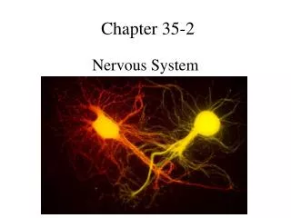
Chapter 35-2. Nervous System. Structure of the Neuron. Neurons come in all shapes & sizes Have common features. Nucleus. Axon terminals. Cell body. Myelin sheath. Dendrites. Axon. Nodes. A Neuron. Nerve Impulses. Resting Neuron – when at rest: - outside of cell = + charge

Chapter 35-2
E N D
Presentation Transcript
Chapter 35-2 Nervous System
Structure of the Neuron Neurons come in all shapes & sizes Have common features Nucleus Axon terminals Cell body Myelin sheath Dendrites Axon Nodes A Neuron
Nerve Impulses • Resting Neuron – when at rest: - outside of cell = + charge - inside of cell = - charge - cell membrane = electrically charged - due to the opposite charges on both sides - caused by the - & + elements crossing the membrane by active transport
Sodium Potassium Pump • Na+/K +pump • How it works: 1. nerve cell membrane pump Na +out of cell & K+ into the cell by active transport 2. as a result: inside – ↑ K+ and ↓ Na+ outside - ↑ Na+ and ↓ K+ 3. the nerve cell membrane allows more K+ to cross the membrane result - K+ goes outside of cell – produces – charge inside cell + charge outside cell
Figure 35-6 Resting Potential Resting Potential Section 35-2 • Electrical charge across the cell membrane of a neuron in its resting state Outside of cell (+ charge) Cell membrane Inside of cell (- charge)
Moving Impulse • A neuron remains resting until it receives a stimulus large enough to start a nerve impulse • An impulse begins when a neuron is stimulated by: 1. another neuron 2. by the environment
Process of Impulses • When it begins, the impulse travels rapidly down the axon, away from cell body towards the axon terminals • This causes the charges to reverse the membrane potential (- to +) - results from the leading edge of impulse opening Na channels which allows Na to flow into the cell - Action Potential – the reversal of charges from – to + - also called “nerve impulse”
Process of an Impulse Section 35-2 Action Potential As the action potential passes, potassium gates open, allowing K+ ions to flow out. At rest. Action Potential Action Potential The action potential continues to move along the axon in the direction of the nerve impulse. At the leading edge of the impulse, the sodium gates open. The membrane becomes more permeable to Na+ ions and an action potential occurs.
Process of Impulses cont. 3. As the action potential passes, gates in the K+ channels open, allowing K+ to flow out - this restores the resting potential - reversing the charge back to normal 4. The axon potential continues to move along the axon until it reaches the organ or gland Nerve impulses are “self-propagating”: - an impulse at any point on the membrane cause an impulse at the next point on the membrane ( row of dominoes)
Threshold • The strength of an impulse is always the same – either there is an impulse or there is not • A stimulus must have enough strength to cause a neuron to start an impulse • The minimum level of a stimulus that is required to activate a neuron is called the THRESHOLD
All or None Principle • Either the stimulus will produce an impulse or it will not produce an impulse. • No in between • If you stack a row of dominoes and push the first one: - either they all fall down or they don’t move at all
Figure 35-8 The Synapse Synapse Section 35-2 • The location at which a neuron can transfer an impulse to another cell • Motor neurons pass impulses to muscle cells Direction of Impulse Dendrite of adjacent neuron Axon Receptor Vesicle Axon terminal Synaptic cleft Neurotransmitter
Figure 35-8 The Synapse Synaptic Cleft Section 35-2 • A space that separates the axon terminal from the dendrites of the next neuron • Contains tiny vesicles filled with neurotransmitters Direction of Impulse Dendrite of adjacent neuron Axon Receptor Vesicle Axon terminal Synaptic cleft Neurotransmitter
Figure 35-8 The Synapse Neurotransmitters Section 35-2 • Chemicals used by a neuron to transmit an impulse across the a synapse to another cell Direction of Impulse Dendrite of adjacent neuron Axon Receptor Vesicle Axon terminal Synaptic cleft Neurotransmitter
How Neurotransmitters Work • When an impulse arrives at the axon terminal, the vesicles release the neurotransmitters into the synaptic cleft • Neurotransmitters diffuse across the cleft and attachment themselves to receptors on the membrane of the next cell • This stimulus cause positive Na to rush across the membrane, stimulating the next cell • If stimulation exceeds the cell’s threshold, a new impulse begins
Life of a Neurotransmitter • Only a fraction of a second after binding to its receptors, the neurotransmitter is released from the membrane. • Neurotransmitters are either: 1. broken down by enzymes 2. taken up & recycled by the axon terminal