Oral vestibule
350 likes | 633 Views
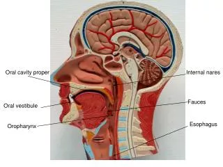
Oral cavity proper. Internal nares. Fauces. Oral vestibule. Esophagus. Oropharynx. Auditory / Pharyngo- Tympanic tube. Cribiform plate of ethmoid. Frontal sinus. Sphenoidal sinus. Nasophayngeal tonsil. Chonchae / turbinates. Soft palate. Palatine tonsil. Uvula. Lingual

Oral vestibule
E N D
Presentation Transcript
Oral cavity proper Internal nares Fauces Oral vestibule Esophagus Oropharynx
Auditory / Pharyngo- Tympanic tube Cribiform plate of ethmoid Frontal sinus Sphenoidal sinus Nasophayngeal tonsil Chonchae / turbinates Soft palate Palatine tonsil Uvula Lingual tonsil Epiglottis Cricoid cartilage
Enamel Crown Dentin Neck Gingiva Cementum Periodontal ligament Alveolar bone Root Root canal with blood vessels and nerves Pulp cavity With blood vessels and nerves
Common bile duct Pancreatic duct Duodenal papillus (major) Plica circulares of duodenum Hepatopancreatic ampulla in the duodenal wall
Gall bladder Spleen Liver Cystic duct Adrenal gland Hepatic duct Kidney Duodenum Head, body and tail of the pancreas
Pancreatic duct Common Bile Duct Plica circulares Spleen Duodenum
Common bile duct Hepatic portal vein Cystic duct Hepatic artery Hepatic duct Round ligament (remnant of umbilical vein) Gall bladder
Lacteal Villi Simple columnar epithelium Crypt of Lieberkuhn Muscularis mucosae Submucosa Circular and Longitudinal layers Of muscularis externa Serosa
Lacteal (green) Villi Lymphatic nodule/ Lymphatic follicle Lamina propria Muscularis mucosae Submucosa with many blood vessels Circular layer of Muscularis externa Longitudinal layer Of muscularis externa
Villi Lacteals Lymph nodule/follicle Crypt of Lieberkuhn Muscularis mucosae Circular and Longitudinal layers of the Muscularis externa
Ulcerative colitis Diverticula Adhesions Irritable Bowel/ Spastic colon Bacterial infection Crohn’s disease (enteritis) Cancer Polyps Appendicitis
Transverse colon Haustra Ascending colon Descending colon Taenia coli Ileocecal valve Sigmoid colon Cecum Rectum
Inner oblique layer of muscle Middle circular layer of muscle Outer longitudinal layer of muscle
Fundus Cardia Pylorus Body
Pylorus Esophagus Site of lower esophageal sphincter/Cardiac sphincter Lesser curvature Pyloric valve/ sphincter Rugae Greater curvature
Lingual tonsil Sublingual gland Submandibular gland
Circumvallate papillae Lingual tonsil Sublingual gland Submandibular gland and duct
Lingual tonsil Sublingual gland Epiglottis Submandibular gland Arytenoid cartilage Cricoid cartilage Trachealis muscle
Enamel Dentin Gingiva Pulp cavity Alveolar bone Root canal Cementum Apical foramen Periodontal ligament
Lacteal Villi Simple columnar epithelium Crypt of Lieberkuhn Muscularis mucosae Lymphatic vessels Goblet cells Circular and Longitudinal layers Of muscularis externa Serosa
Submandibular duct (Wharton) Sublingual ducts (Rhivinus) Parotid gland Parotid duct / Stenson’s duct Sublingual gland Submandibular gland
Lesser omentum Esophagus Pylorus Fundus Body Greater omentum Cardia
Low magnification of numerous filliform papillae on surface of tongue
Numerous Filliform Papillae (note lack of taste buds) Skeletal Muscle of Tongue
Foliate or Fungiform Papillae Taste Buds
Taste Buds Stratified squamous epithelium Foliate or Fungiform Papillae on Tongue
Gustatory Pore Gustatory Cells In Taste Bud Taste Buds Stratified squamous epithelium
Gustatory pore with free nerve endings Nuclei of Gustatory Cells Magnified Taste Buds
Liver Central veins in center of Hexagonal lobes Portal triads
Liver Portal triad containing branches of hepatic portal vein, hepatic artery and bile duct Hepatocytes
Numerous Peyer’s Patches Duodenum Jejenum Ileum
Crypts of Lieberkuhn Villi Muscularis mucosae Brunner’s or Submucosal glands Muscularis externa, Circular layer
Muscularis Mucosa Submucosa Villi Lumen of GI Tract Outer Longitudinal Layer Inner Circular Layer of the Muscularis Externa Peyer’s Patches in the submucosa Of the ileum
Peyer’s Patches in the submucosa Muscularis (externa) Submucosa Ileum Villi Lumen
Parotid gland Parotid duct / Stenson’s duct Sublingual gland Submandibular gland