Download
1 / 131
1.38k likes | 1.98k Views
7 The Axial Skeleton. An Introduction to the Axial Skeleton. Learning Outcomes 7-1 Identify the bones of the axial skeleton, and specify their functions.

E N D
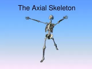
7 The Axial Skeleton
An Introduction to the Axial Skeleton • Learning Outcomes • 7-1 Identify the bones of the axial skeleton, and specify their functions. • 7-2 Identify the bones of the cranium and face, and explain the significance of the markings on the individual bones. • 7-3 Identify the foramina and fissures of the skull, and cite the major structures using the passageways. • 7-4 Describe the structure and functions of the orbital complex, nasal complex, and paranasal sinuses.
An Introduction to the Axial Skeleton • Learning Outcomes • 7-5 Describe the key structural differences among the skulls of infants, children, and adults. • 7-6 Identify and describe the curvatures of the spinal column, and indicate the function of each. • 7-7 Identify the vertebral regions, and describe the distinctive structural and functional characteristics of vertebrae in each region. • 7-8 Explain the significance of the articulations between the thoracic vertebrae and the ribs, and between the ribs and sternum.
An Introduction to the Axial Skeleton • Structures of Bones • Articulations • Contacts with other bones • Landmarks (bone markings; marks) • Areas of muscle and ligament attachment • Foramina • Openings for nerves and blood vessels
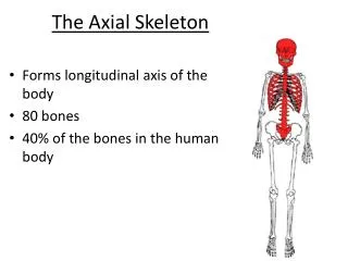
7-1 The Axial Skeleton • The Axial Skeleton • Forms the longitudinal axis of the body • Has 80 bones • The skull • 8 cranial bones • 14 facial bones • Bones associated with the skull • 6 auditory ossicles • The hyoid bone
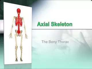
7-1 The Axial Skeleton • The Axial Skeleton • The vertebral column • 24 vertebrae (singular = vertebra) • The sacrum • The coccyx • The thoracic cage • 24 ribs • The sternum
Figure 7-1a The Axial Skeleton SKELETAL SYSTEM 206 80 APPENDICULAR SKELETON AXIAL SKELETON (see Figure 8–1) Cranium 8 Skull Face 14 Skull and associated bones 29 Auditory ossicles 6 Associated bones 1 Hyoid Sternum 1 Thoracic cage 25 Ribs 24 Vertebrae 24 Vertebral column 1 Sacrum 26 1 Coccyx An anterior view of the entire skeleton, with the axial components highlighted. The numbers in the boxes indicate the number of bones in the adult skeleton.
Figure 7-1b The Axial Skeleton Skull Cervical vertebrae Sternum Thoracic vertebrae Ribs Costal cartilages Lumbar vertebrae Sacrum Coccyx Anterior (left) and posterior (right) views of the axial skeleton. The individual bones associated with the skull are not visible.
7-1 The Axial Skeleton • Functions of the Axial Skeleton • Supports and protects organs in body cavities • Attaches to muscles of head, neck, and trunk • Performs respiratory movements • Stabilizes parts of appendicular skeleton
7-2 The Skull • The Skull • Protects: • The brain • Entrances to respiratory system • Entrance to digestive system • Contains: • 22 bones • 8 cranial bones • Form the braincase or cranium • 14 facial bones • Protect and support entrances to digestive and respiratory tracts
Figure 7-2 Cranial and Facial Subdivisions of the Skull SKULL CRANIUM FACE 14 ASSOCIATED BONES 8 7 2 Maxillary bones 1 Occipital bone Palatine bones 2 Parietal bones 2 1 Hyoid bone Auditory ossicles enclosed in temporal bones (detailed in Chapter 17) Nasal bones 1 Frontal bone 2 6 Temporal bones 2 Inferior nasal conchae 2 Sphenoid 1 2 Zygomatic bones Ethmoid 1 2 Lacrimal bones 1 Vomer 1 Mandible
Figure 7-2 Cranial and Facial Subdivisions of the Skull PARIETAL BONE FRONTAL BONE TEMPORAL BONE SPHENOID ETHMOID OCCIPITAL BONE LACRIMAL BONE NASAL BONE Cranial bones VOMER ZYGOMATIC BONE MAXILLA MANDIBLE Facial bones
7-2 The Skull • Cranial Bones • Enclose the cranial cavity • Which contains the brain • And its fluids, blood vessels, nerves, and membranes
7-2 The Skull • Facial Bones • Superficial facial bones • For muscle attachment • Deep facial bones • Separate the oral and nasal cavities • Form the nasal septum
Figure 7-3a The Adult Skull Sagittal suture PARIETAL BONE (right) PARIETAL BONE (left) Lambdoid suture OCCIPITAL BONE Squamous suture TEMPORAL BONE Mastoid process Styloid process Occipital condyle External occipital protuberance MANDIBLE Posterior view
Figure 7-3b The Adult Skull OCCIPITAL BONE Lambdoid suture PARIETAL BONE (right) PARIETAL BONE (left) Sagittal suture Coronal suture FRONTAL BONE ZYGOMATIC BONE NASAL BONES Superior view
Figure 7-3c The Adult Skull Coronal suture FRONTAL BONE PARIETAL BONE SPHENOID Supra-orbital foramen Squamous suture TEMPORAL BONE NASAL BONE LACRIMAL BONE Squamous part of temporal bone ETHMOID Lambdoid suture Infra-orbital foramen OCCIPITAL BONE MAXILLA External acoustic meatus ZYGOMATIC BONE Mastoid process Styloid process MANDIBLE Zygomatic process of temporal bone Mental foramen Zygomatic arch Temporal process of zygomatic bone Mental protuberance Lateral view
Figure 7-3d The Adult Skull Coronal suture PARIETAL BONE FRONTAL BONE SPHENOID TEMPORAL BONE Supra-orbital foramen ETHMOID Optic canal PALATINE BONE Superior orbital fissure LACRIMAL BONE Inferior orbital fissure Temporal process of zygomatic bone ZYGOMATIC BONE Mastoid process of temporal bone Infra-orbital foramen NASAL BONE Middle nasal concha (part of ethmoid) MAXILLA Perpendicular plate of ethmoid INFERIOR NASAL CONCHA Bony nasal septum VOMER MANDIBLE Mental protuberance Anterior view Mental foramen
Figure 7-3e The Adult Skull FRONTAL BONE MAXILLA ZYGOMATIC BONE VOMER SPHENOID PALATINE BONE Foreman ovale Zygomatic arch Medial and lateral pterygoid processes Styloid process Mandibular fossa Foramen lacerum Carotid canal External acoustic meatus TEMPORAL BONE Mastoid process Jugular foramen Stylomastoid foramen Lambdoid suture Occipital condyle OCCIPITAL BONE Foramen magnum External occipital protuberance Inferior view
Figure 7-4a The Sectional Anatomy of the Skull Coronal suture PARIETAL BONE FRONTAL BONE SPHENOID Squamous suture Sphenoidal sinus (right) TEMPORAL BONE Frontal sinus Crista galli Lambdoid suture NASAL BONE ETHMOID Hypophyseal fossa of sella turcica VOMER Internal acoustic meatus PALATINE BONE OCCIPITAL BONE MAXILLA Hypoglossal canal Styloid process MANDIBLE Medial view of a sagittal section through the skull.
Figure 7-4b The Sectional Anatomy of the Skull FRONTAL BONE Crista galli ETHMOID Cribriform plate Sella turcica Foramen rotundum SPHENOID Foramen lacerum Foramen ovale Foramen spinosum TEMPORAL BONE Carotid canal Internal acoustic meatus Foramen magnum PARIETAL BONE Jugular foramen Internal occipital crest Hypoglossal canal OCCIPITAL BONE Superior view of a horizontal section through the skull, showing the floor of the cranial cavity. Compare with part (a) and with Figure 7–3e.
7-2 The Skull • Superficial Facial Bones • Maxillae = maxillary bones • Lacrimal • Nasal • Zygomatic • Mandible • Deep Facial Bones • Palatine • Inferior nasal conchae • Vomer
7-2 The Skull • Sinuses • Cavities that decrease the weight of the skull • Lined with mucous membranes • Protect the entrances of the respiratory system
7-2 The Skull • Sutures • The immovable joints of the skull • The four major sutures • Lambdoid suture • Coronal suture • Sagittal suture • Squamous suture
7-2 The Skull • Lambdoid Suture • Separates occipital from parietal bones • May contain sutural (Wormian) bones • Coronal Suture • Attaches frontal bone to parietal bones • The calvaria (skullcap) • Consists of occipital, parietal, and frontal bones
7-2 The Skull • Sagittal Suture • Between the parietal bones • From lambdoid suture to coronal suture • Squamous Sutures • Form boundaries between temporal bones and parietal bones
7-3 The Cranial Bones of the Skull • The Cranial Bones • Occipital bone • Parietal bones • Frontal bone • Temporal bones • Sphenoid • Ethmoid
7-3 The Cranial Bones of the Skull • The Occipital Bone • Functions of the occipital bone • Forms the posterior and inferior surfaces of the cranium • Articulations of the occipital bone • Parietal bones • Temporal bones • Sphenoid • First cervical vertebra (atlas)
7-3 The Cranial Bones of the Skull • TheOccipital Bone • Marks of the occipital bone • External occipital protuberance • External occipital crest • Occipital condyles articulate with neck • Inferior andsuperior nuchal lines: attachment site of muscles and ligaments
7-3 The Cranial Bones of the Skull • TheOccipital Bone • Foramina of the occipital bone • Foramen magnum connects cranial and spinal cavities • Jugular foramen for jugular vein • Hypoglossal canals for hypoglossal nerves
Figure 7-5a The Occipital and Parietal Bones Hypoglossal canal Occipital condyle Foramen magnum External occipital crest Inferior nuchal line Superior nuchal line External occipital protuberance Occipital bone, inferior view
7-3 The Cranial Bones of the Skull • The Parietal Bones • Functions of the parietal bones • Form part of the superior and lateral surfaces of the cranium • Articulations of the parietal bones • Other parietal bone • Occipital bone • Temporal bone • Frontal bone • Sphenoid
7-3 The Cranial Bones of the Skull • The Parietal Bones • Marks of the parietal bones • Superior and inferiortemporal lines • To attach temporalis muscle • Grooves for cranial blood vessels
Figure 7-5b The Occipital and Parietal Bones Superior temporal line Inferior temporal line Right parietal bone, lateral view
7-3 The Cranial Bones of the Skull • The Frontal Bone • Functions of the frontal bone • Forms the anterior cranium and upper eye sockets • Contains frontal sinuses • Articulations of the frontal bone • Parietal bone • Maxillary • Metopic suture • Ethmoid • Lacrimal bone • Zygomatic bone • Sphenoid • Nasal bone
7-3 The Cranial Bones of the Skull • The Frontal Bone • Marks of the frontal bone • Frontal squama (forehead) • Supra-orbital margin (protects eye) • Lacrimal fossa (for tear ducts) • Frontal sinuses
7-3 The Cranial Bones of the Skull The Frontal Bone Foramina of the frontal bone Supra-orbital foramen For blood vessels of eyebrows, eyelids, and frontal sinuses Supra-orbital notch An incomplete supra-orbital foramen
Figure 7-6a The Frontal Bone Frontal (metopic) suture Frontal squama Superior temporal line Supra-orbital notch Supra-orbital margin Anterior surface
Figure 7-6b The Frontal Bone Frontal sinus Supra-orbital foramen Supra-orbital margin Lacrimal fossa Inferior (orbital) surface
7-3 The Cranial Bones of the Skull • The Temporal Bones • Functions of the temporal bones • Part of lateral walls of cranium and zygomatic arches • Articulate with mandible • Surround and protect inner ear • Attach muscles of jaws and head
7-3 The Cranial Bones of the Skull • The Temporal Bones • Articulations of the temporal bones • Zygomatic bone • Sphenoid • Parietal bone • Occipital bone • Mandible
7-3 The Cranial Bones of the Skull • The Temporal Bones • Marks of the temporal bones • Squamous part • Zygomatic process • Mandibular fossa • Mastoid process • Styloid process • Petrous part • Auditory ossicles
7-3 The Cranial Bones of the Skull Squamous Part Borders the squamous suture Zygomatic Process Inferior to the squamous portion Articulates with temporal process of zygomatic bone Forms zygomatic arch (cheekbone) Mandibular Fossa Articulates with the mandible
7-3 The Cranial Bones of the Skull • Mastoid Process • For muscle attachment • Contains mastoid air cells connected to middle ear • Styloid Process • To attach tendons and ligaments of the hyoid, tongue, and pharynx • Petrous Part • Encloses structures of the inner ear
7-3 The Cranial Bones of the Skull • Auditory Ossicles • Three tiny bones in tympanic cavity (middle ear) • Transfer sound from tympanic membrane (eardrum) to inner ear
7-3 The Cranial Bones of the Skull • The Temporal Bones • Foramina of the temporal bones • Carotid canal for internal carotid artery • Foramen lacerum • For carotid and small arteries • Hyaline cartilage • Auditory tube
7-3 The Cranial Bones of the Skull • The Temporal Bones • Foramina of the temporal bones • External acoustic meatus (canal) ends at tympanic membrane • Stylomastoid foramen for facial nerve • Internal acoustic meatus (canal) • For blood vessels and nerves of the inner ear • Facial nerve
Figure 7-7a The Temporal Bones Petrous part Squamous part Internal acoustic meatus Mastoid process Zygomatic process Styloid process Medial view of the right temporal bone
Figure 7-7b The Temporal Bones Squamous part Mandibular fossa External acoustic meatus Zygomatic process Styloid process Mastoid process Lateral view of the right temporal bone
Figure 7-7c The Temporal Bones External acoustic meatus Mastoid process, cut to show mastoid air cells A cutaway view of the mastoid air cells