Download
1 / 22
230 likes | 344 Views
Delve into the world of thrombosis with Dr. Nisreen Abu Shahin as she explains the pathogenesis and morphology of blood clots. Learn about endothelial injury, stasis, hypercoagulability, and more. Discover how thrombi form, their locations, and potential consequences. Stay informed on factors contributing to thrombosis and risk management strategies.

E N D
Thrombosis Dr. Nisreen Abu Shahin Assistant Professor of Pathology Pathology Department University of Jordan
Artery (A) vsvien (V) The thickness of the tunica media is larger in arteries than in veins because blood pressure inside them is higher and to pump blood to down stream tissues (the tissues supplied by this artery)
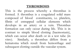
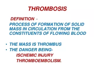
Thrombosis ** the doc. read everything in the slides A Some of them might result in the activation of the other and they are often found in combinations. • Pathogenesis (called Virchow's triad): • Endothelial* Injury ( Heart, Arteries) • Stasis (abnormal blood flow) • Blood Hypercoagulability * Endothelial cells are special type of cells that cover the inside surface of blood vessels and heart (including the valves). -They are not a type of blood cells. -Their function (when healthy and not injured) is protection against thrombosis by secreting and controlling certain mediators that prevent the formation of blood clot and when formed they help to lyse it
Contribution of Endothelial Cells to Coagulation • Intact endothelial cells maintain liquid blood flow by: 1- inhibiting platelet adherence 2- preventing coagulation factor activation 3- lysing blood clots that may form. • Endothelial cells can be stimulated by direct injury or by various cytokines that are produced during inflammation. • Endothelial injury results in: 1- expression of procoagulant proteins (tissue factor and vWF)local thrombus formation. 2- exposure of underlying vWF and basement membrane collagen platelet aggregation and thrombus formation. Endothelial = endothelial stimulation injury
Response of Vascular Wall Cells to Injury After any tissue damage inflammation and then healing will occur • Injury to the vessel wall results in a healing response, involving: 1- Intimal expansion by proliferating SMCs and newly synthesized ECM. 2- The recruitment and activation of the SMCs in which involves signals from cells (e.g., ECs, platelets, and macrophages), as well as mediators derived from coagulation and complement cascades. • Pathologic effect of vascular healing Excessive thickening of the intima luminal stenosis & blockage of vascular flow
Narrowing of the lumen Thicken-ing of the vessel wall Normal blood vessel
Mainly caused by Physical injury Induction of inflammation • Causes of Endothelial injury • Valvulitis(inflammation of heart valves) • MI (myocardial infarction) • Atherosclerosis • Traumatic or inflammatory conditions • Increased Blood Pressure • Endotoxins (LPS found in the cell wall of gram –ve bacteria) • Hypercholesterolemia (the doc. Says it acts as an antigorus body :/). Might cause atherosclerosis then thrombosis ? • Radiation • Smoking Physical injury
Center Periphery Platelets - RBCS-WBCS- clear zone Easier to occur in veins because of the lower musculature • Stasis • Stasis is a major factor in venous thrombi • Normal blood flow is laminar (platelets flow centrally in the vessel lumen, separated from the • endothelium by a slower moving • clear zone of plasma) • Stasis and turbulence cause the followings: In the case of stasis this arrangement is disrupted, and platelets come in contact with the endothelial cells and clot forms Meaning that the clotting factors will work for a longer time Stasis results in hypoxia caused by the decreased oxygen tension --> endothelial injury
Causes of Stasis • Atherosclerosis • Aneurysms (enlargement of an artery) • Myocardial Infarction ( Non-cotractile fibers) • Mitral valve stenosis (atrial dilation) • Hyper viscosity syndrome (PCV and Sickle Cell anemia)
Mutations In clotting factors ( have increased activity) or clotting factors inhibitors(have decreased activity) • Hypercoagulability A. Genetic (primary): • mutations in the factor V gene and the prothrombin gene are the most common B. Acquired (secondary): • multifactorial and is therefore more complicated • causes include: Immobilization, MI, AF, surgery, fractures, burns, Cancer, Prosthetic cardiac valves …etc causes Might be due to thrombus formation when they metastasize, Or clotting factors production (paraneoplastic syndrome)
Morphology of thrombi • Can develop anywhere in the CVS (e.g., in cardiac chambers, valves, arteries, veins, or capillaries). • Arterial or cardiac thrombi begin at sites of endothelial injury or turbulence; and are usually superimposed on an atherosclerotic plaque • Venous thrombi occur at sites of stasis. Most commonly the veins of the lower extremities (90%) • Thrombi are focally attached to the underlying vascular surface. • The propagating portion of a thrombus is poorly attached fragmentation and embolus formation
Read this after getting through organization and recanalization in slide 21 ARTERY WITH AN OLD THROMBUS. A, H&e-stain. B, Stain For Elastic Tissue. The Original Lumen Is Delineated By The Internal Elastic Lamina (Arrows) And Is Totally Filled With Organized Thrombus, Now Punctuated By A Number Of Recanalized Channels (White Spaces).
lines of Zahn • Thrombi can have grossly (and microscopically) apparent laminations called lines of Zahn; these represent pale platelet and fibrin layers alternating with darker erythrocyte-rich layers. • Such lines are significant in that they represent thrombosis of flowing blood (can potentially distinguish antemortem thrombosis from postmortem clots) • postmortem blood clots are bland non-laminated clots (no lines of Zahn) They appear in antemortem blood clots, and only used to differentiae between ante- and post-mortem clots in forensics
Mural thrombi • Thrombi occurring in heart chambers or in the aortic lumen. • Causes include: Abnormal myocardial contraction (e.g. arrhythmias, dilated cardiomyopathy, or MI) or endomyocardial injury (caused by myocarditis, catheter trauma) • Vegetations • Thrombi on heart valves are called vegetations: Types: 1- infectious (Bacterial or fungal blood-borne infections)(e.g. infective endocarditis,). 2-Non-bacterial thrombotic endocarditisoccur on sterile valves.
Fate of thrombi • Propagation accumulate additional platelets and fibrin, eventually causing vesselobstruction leading to hypoxia and necrosis • Embolization Thrombi dislodge or fragment and are transported elsewhere in the vasculature • Dissolution Thrombi are removed by fibrinolytic activity (only in recent thrombi) the ones formed in less than 5 hours • Organization* and recanalization Thrombi induce inflammation and fibrosis. These can recanalize (re-establishing some degree of flow), • or they can be incorporated into a thickened vessel wall *Organization refers to the ingrowth of endothelial cells, smooth cells and fibroblasts into the fibrin rich thrombus. 5. Superimposed infection (Mycoticaneurysm) Less compared to the other fates 1 1 Capillary sized channels in the thrombus 2 thrombus 2 Thrombus incorporated into the vessel wall
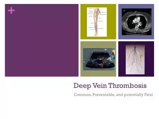
Venous thrombi • (veins of the legs) are most common • Superficial: e.g. Saphenous veins. - can cause local congestion, swelling, pain, and tenderness along the course of the involved vein, but they rarely embolize • Deep: e.g. Popliteal, Femoral and iliac vein. • more serious because they may embolize • can occur with stasis or in a variety of hypercoagulable states