Download
1 / 39
430 likes | 520 Views
This article discusses the surgical management of lung infections, specifically focusing on empyema and lung abscesses. It covers definitions, causes, clinical presentations, diagnosis, and management strategies for both conditions. The etiology, bacteriology, clinical presentation, diagnosis methods, and treatment options for empyema are detailed, including thoracentesis, CT scans, and surgical interventions such as chest tube insertion and thoracotomy. For lung abscesses, the definition, types, risk factors, bacteriology, diagnosis methods like CT scans and bronchoscopy, and management principles involving antibiotic therapy and drainage are explained.

E N D
EMPYEMA LUNG ABSCESS(SURGICAL LUNG INFECTIONS) Dr. Mahmoud ABU-ABEELEH The University of JORDAN. Faculty of Medicine. 4-10-2016
ANATOMY. • Empyema Definition classification causes diagnosis management indications for surgery • Lung abscesses definition causes clinical presentations diagnosis management
EMPYEMA • Definition:Invading of the pleural space with bacteria which result in accumulation of pus . • Classification :(American Thoracic Society) • Stage 1 :Exudative , with swelling of the pleural membranes as a result of ↑ permeability of swollen membranes(Uncomplicated Acute stage) • Stage 2:Fibropurulent(Transitional)with heavy fibrin deposits. • Stage 3:Organizing or Chronic phase.With ingrowth of fiboblast and deposition of collagen
ETIOLOGY: • PARAPNEUMONIC(secodary to a pneumonia)the most common • Post trauma. • Post surgery(esophageal or pulmonary( • Subphrenic Abscess
Bacteriology • :Before ABO 10% of Pts survived pneumonia developed EMPYEMA(Streptococci & Pneumococci are the most freuquent) • After ABO the incidence as well as the mortality↓. Staph become more prevelent ,90% of empyema in children .
Incidence of Empyema according to Bacteria causing pneumonia
Clinical presentation • Pleuritic chest pain ,fever, S.O.B ,Tacycardia AS Pneumonia.If prolonged symptoms SUSPECT EMPYEMA. • Anaerobic :indolent • P/E:Toxic anxious pt,tacycardia,tacypnea,restricted chest wall excursion,↓ air entry,dullness on percussion. • Chronic ptClubbing,Anaemia,wt loss.
DIAGNOSIS: • CBC:↑ WBCwith shift to left,↑ CRP ESR. • CXR:Effusion,↑thickness of the pleura, Air fluid level. • THORACOCENTESIS: • Empyema fluid • PH <7.0 • Glucose < 40 mg/dLLDH > 1000 IU/dLPositive Gram stainPositive culture (50%)Specific gravity > 1.018WBC > 500 cells/mm3Protein > 2.5 g/dL
CT Scan: • Localize collection. • Identify the underlying parenchymal disease,. • Distinguish it from lung abscess. • Fluid density,loculations. • Therapeutic:CT-guided aspiration.
Managment • Antibiotics.3rd generation cephalosporine,clindamycin till the result of G stain ,C&S. • Evacuation of pus from the pleural space. In stage 1 thoracocentesis,other wise Chest tube insertion • Obliteration of the empyema cavity.
Chest Tube Insertion • Procedure • local anaesthesia • Scrubing & draping • An incision is made along the upper border of the rib • By a curved clamp the track is developed by blunt dissection splitting the fibres. A track developed with the operator's finger • The clamp is angled over the rib & dissection continued until pleura is entered.
Chest Tube Insertion • Procedure • A large-bore (32 or 36F) chest tube is passed into the pleural cavity. • The tube is connected to an underwater seal and sutured / secured in place.a U-stitch • A chest X-ray is taken to confirm placement & position.
Clinical improvement within 48 hrs. • ≥80% of stage 1 managed conservatively. • Stage 3 80% require thoracotomy. • Intrapleural Fibrinolytic therapy; STK or Urokinase to break loculations produced by membranes composed of fibrin. • V.A.T .S. • THORACOTOMY: decortication.
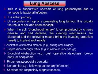
LUNG ABSCESS • Definition:Sub acute pulmonary infection in which the chest X ray shows cavity within the lung parenchyma. • Before ABO era ,high mortality,. • ACUTE &CHRONIC:if duration< 6 weeks. • PRIMARY &SECONDARY • PRIMARY: • Aspiration:The most frequent . • Post-Pneumonic
Secondary: • Obstructing carcinoma. • COPD • Metastatic from extrathoracic source septisemia. • F.B aspiration. • Pulmonary infarctions. • The individuals with high risk: ALCOHOL ABUSE, hx of Aspiration,Old TB, Epilepsy,drugabuse,COPD. • In endemic areas TB:20% of lung abscesses have TB.
BACTERIOLOGY: • ANAEROBES:75-80% • Bacteroidfragilis. • Fusibacterium bacilli. • Peptostreptococci. • Provetella. • AEROBIC: • Kleibsiella &Pseudomonus:IN obstructive infections &Nasocomial. • Staph.Auereus. • S. pneumonia • H.influenza.
DIAGNOSIS: • Symptoms:Fever intermittent &night sweats chills.Purulent Foul-smelling sputum is highly suggestive. • Hx of Aspiration,.Sepsis→Respiratory failure. • Signs:Tachpnea,consolidation,local chest wall tenderness. • CXR: • Pneumonitis pattern early→Air-fluid level.
SPUTUM analysis&cultureAerobic,anaerobic,fungal &TB. • CT-scan. • FibroopticBronchoscopy:is mandatory • Take samples for culture. • R/O endobronchialtumour or obstruction. • To assess if can be drained internally.
SITES: • Superior segment of Rt lower lobe. • Lat. Part of Post. Segment of R.U.L. • Superior segment of L.L.L. • D.Dx of cavitary lung lesion: • Cavitary carcinoma. • T.B or fungal abscess. • Pyogenic lung abscess. • Empyema with bronchopleural fistula.
Managment:Principles of therapy: • Identify the organism→proper ABO therapy for 6-8 wks. • Drainage: • Chest physiotherapy. • Bronchoscopy=internal darainaige or indwelling transbronchial catheter drainage. • Percutaneous cath. Drainage. • SURGERY. • 80-90%of Lung abscess respond to medical tt.Flagyl or Clindamycinfor anaerobes. • Gentamicin or 3rd generation cephalosporines for aerobes.
External drainage: • If remain septic. • Failure to wean from mechanical ventilation. • Soiling of the contralateral lung. • Abscess cavity >4 cm& under tension on CXR. • ↑ size while on ABO. • Chest tube thoracostom. • CT-guided catheter. • Open pneumonostomy =MONALDI procedure. • 30% of Pt will need definitive surgery. • Clinical improvement within 48 hrs.
INDICATIONS FOR SURGERY: • Acute : for complications • Bronchopleural fistula. • Empyema. • Hemoptysis.(Massive) • Chronic =Definitive. • Persistant symtoms despite long term ABO therapy. • Suspecius of carcinoma. • Complications:Empyema,bronchopleural fistula. • Persistant cavity >6 cm after ABO therapy.
Lobectomy is the standard procedure. • Mortality: • 2.5% after community acquired pneumonia. • 66% with Nasocomial infections. • Underlying diseases. • Size of the abscess >6 cm. • Organism: Pseudomonus & G –ve the highest.
QUESTIONS SUMMARY