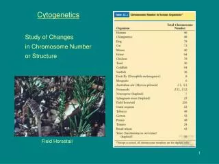

Advances in Cytogenetics: Understanding Chromosomal Abnormalities
E N D
Presentation Transcript
Part 1 Cytogenetics Dr. Mohammed Hussein M.B.Ch.B, MSC, PhD, DCH (UK), MRCPCH
Introduction • Numerical Chromosomal Abnormalities • Structural Chromosomal Abnormalities • Advances in Molecular Cytogenetics
Chromosome abnormalities are: • Seen in 1 /150 live births • The leading known cause of mental retardation • The leading known cause of pregnancy loss • 50% of fetal losses during the first trimester of pregnancy • 20% of fetuses lost during the second trimester of pregnancy
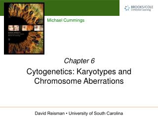
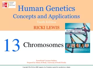
Karyotyping • Chromosomes are most easily visualized during the metaphase stage of mitosis, when they are maximally condensed • They are photographed under the microscope to create a karyotype • Karyotype is an ordered display of the 23 pairs of human chromosomes in a typical somatic cell
Karyogram • Karyogramrepresents a drawing of each type of chromosome • The presentation is haploid (only one copy of each chromosome is shown)
Chromosome Banding • To visualize chromosomes in a karyotype, various stains are applied so that banding is evident • G-banding:Mitotic chromosomes are partially digested with trypsin (to digest some associated protein) and then stained with Giemsa, a dye that binds DNA
p X .1 2 2 X q two-two point one NOTX q twenty-two point one
Metacentric chromosomes: have the centromere near the middle • Submetacentric chromosomes: have the centromere displaced toward one end • Acrocentric chromosomes: have the centromere far toward one end
Acrocentric Chromosomes • Have the centromere far toward one end • Chromosomes 13, 14,15, 21, and 22 • Only the acrocentric chromosomes are involved in Robertsonian translocations
Mnemonic • Acrocentric chromosomes • 1,2,3,4,5 • 21, 22, 13 , 14 , 15
46,XY Normal male • 46,XX Normal female • 47,XY,+21 Male with extra chromosome no.21 (Trisomy 21)(Down syndrome) • 47,XX,+13 Female with extra chromosome no.13 (Trisomy 13)(Patau syndrome) • 45,XX,–13 Female with missing chromosome no.13 (Monosomy 13) • 47,XX,+18 Female with extra chromosome no.18 (Trisomy 18)(Edward syndrome) • 47,XXY Male with extra X chromosome (Klinefelter syndrome) • 45,X Female with missing X chromosome (Turner syndrome) • 46,XY,t(2p;8p) Male with translocation between short arms of chromosomes 2 & 8 • 46,XY, del(5p) Male with deletion in short arm of chromosome 5 (Cri Du Chat syndrome) • 46,X,r(X) Female with ring chromosomes X (Turner syndrome)
Chromosome Abnormalities Numerical Chromosomal Abnormalities Structural Chromosomal Abnormalities
Numerical Chromosome Abnormalities • Polidy : is the no. of the sets of chromosomes in the cell • Euploid: When a cell has a multiple of 23 chromosomes, it is said to be euploid. • Haploid: 1 set of 23 chromosome (gametes) • Diploid: 2 sets of 23 chromosomes (46) (somatic cells) • Triploidy: 3 sets of 23 chromosomes (69) • Tetraploidy: 4 sets of 23 chromosomes (92)
Triploidy • Result of the fertilization of an ovum by two sperm cells • Is common at conception, but the vast majority of these conceptions are lost prenatally • However, about 1 in 10,000 live births is a triploid • These babies have multiple defects of the heart and central nervous system, and they do not survive
Tetraploidy • This lethal condition is much rarer than triploidy among live births • Only a few cases have been described
Aneuploidy • Aneuploidy, a deviation from the euploid number, represents the gain (+) or loss (–) of a specific chromosome • Two major forms of aneuploidy are observed: • Monosomy (loss of a chromosome) (–) • Trisomy (gain of a chromosome) (+)
Aneuploidy • Autosomal aneuploidy • Monosomy (–) • Trisomy (+) • Sex chromosome aneuploidy • Monosomy (–) • Trisomy (+)
Autosomal aneuploidy • All autosomal monosomies are inconsistent with a live birth • Only three autosomal trisomiesare consistent with a live birth • Trisomy 13 • Trisomy 18 • Trisomy 21
Trisomy 13 (Patau Syndrome) 47 ,XX ,+13 • 47,XY,+13 or 47,XX,+13
Trisomy 18 (Edward Syndrome) 47 ,XY ,+18 • 47,XY,+18 or 47,XX,+18
Trisomy 21 (Down Syndrome) 47 ,XX ,+21 • 47,XY,+21 or 47,XX,+21
Sex chromosome aneuploidy • Relatively common • Have less severe consequences than does autosomal aneuploidy.
Some generalizations are helpful • If a Y chromosome is present, the phenotype is male (regardless the no. of X ) • 46,XY = Male • 46,XX = Female • 45,XO = Female (Turner) • 47,XXY = Male ( Klinefelter)
Some generalizations are helpful • One X chromosome is required for survival and any other X will become a Barr body • 46,XX = • One Bar body • 46,XY = • No Bar body • 45,XO = • No Bar body • 47,XXY = • One Bar body • 47,XXX = • Tow Bar bodies
Sex chromosome aneuploidies • Extra X chromosome: • Male with extra X = 47,XXY • Female with extra X = 47,XXX • Extra Y chromosome • Male with extra Y = 47,XYY • Missing X chromosome: • Female with missing X = 45,X or45,XO
Klinefelter Syndrome 47 ,XXY
Turner Syndrome 45 ,X or 45,XO
Triple X Syndrome 47 ,XXX