Download
1 / 46
500 likes | 907 Views
Introduction to Tissue culture. Sompol Tapechum M.D., Ph.D. Department of Physiology Faculty of Medicine Siriraj Hospital. Objectives. After the session, students should be able to explain the meaning of tissue culture and various types of tissue culture the application of tissue culture

E N D
Introduction to Tissue culture Sompol Tapechum M.D., Ph.D. Department of Physiology Faculty of Medicine Siriraj Hospital
Objectives • After the session, students should be able to explain • the meaning of tissue culture and various types of tissue culture • the application of tissue culture • the advantages and disadvantages of each type of tissue culture • the significant of culture environment on tissue culture • the basic procedure of tissue culture • the safety consideration for tissue culture work Experimental Methods 2006
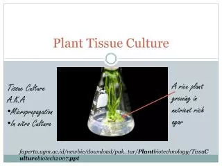
What is tissue culture? • In vitro culture (maintain and/or proliferate) of cells, tissues or organs • Types of tissue culture • Organ culture • Tissue culture • Cell culture Experimental Methods 2006
Organ culture • The entire embryos or organs are excised from the body and culture • Advantages • Normal physiological functions are maintained. • Cells remain fully differentiated. • Disadvantages • Scale-up is not recommended. • Growth is slow. • Fresh explantation is required for every experiment. Experimental Methods 2006
Tissue Culture • Fragments of excised tissue are grown in culture media • Advantages • Some normal functions may be maintained. • Better than organ culture for scale-up but not ideal. • Disadvantages • Original organization of tissue is lost. Experimental Methods 2006
Cell Culture • Tissue from an explant is dispersed, mostly enzymatically, into a cell suspension which may then be cultured as a monolayer or suspension culture. • Advantages • Development of a cell line over several generations • Scale-up is possible • Disadvantages • Cells may lose some differentiated characteristics. Experimental Methods 2006
EMP04 7
Why do we need Cell culture? • Research • To overcome problems in studying cellular behavior such as: • confounding effects of the surrounding tissues • variations that might arise in animals under experimental stress • Reduce animal use • Commercial or large-scale production • Production of cell material: vaccine, antibody, hormone Experimental Methods 2006
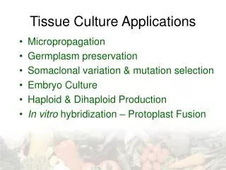
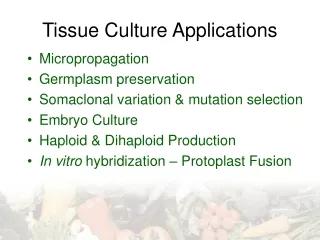
Cell culture application Experimental Methods 2006
Advantages of Cell culture • Advantages: • Absolute control of physical environment • Homogeneity of sample • Less compound needed than in animal models • Disadvantages: • Hard to maintain • Only grow small amount of tissue at high cost • Dedifferentiation • Instability, aneuploidy Experimental Methods 2006
Types of Cell culture • Primary Cultures • Derived directly from excised tissue and cultured either as • Outgrowth of excised tissue in culture • Dissociation into single cells (by enzymatic digestion or mechanical dispersion) • Advantages: • usually retain many of the differentiated characteristics of the cell in vivo • Disadvantages: • initially heterogeneous but later become dominated by fibroblasts. • the preparation of primary cultures is labor intensive • can be maintained in vitro only for a limited period of time.
Types of Cell culture • Continuous Cultures • derived from subculture (or passage, or transfer) of primary culture • Subculture = the process of dispersion and re-culture the cells after they have increased to occupy all of the available substrate in the culture • usually comprised of a single cell type • can be serially propagated in culture for several passages • There are two types of continuous cultures • Cell lines • Continuous cell lines
Types of continuous culture • Cell lines • finite life, senesce after approximately thirty cycles of division • usually diploid and maintain some degree of differentiation. • it is essential to establish a system of Master and Working banks in order to maintain such lines for long periods Experimental Methods 2006
Types of continuous culture • Continuous cell lines • can be propagated indefinitely • generally have this ability because they have been transformed • tumor cells. • viral oncogenes • chemical treatments. • the disadvantage of having retained very little of the original in vivo characteristics Experimental Methods 2006
Transformation VS Transfection • Transformation • Spontaneous or induced permanent phenotypic changes resulting from change in DNA and gene expression • growth rate • mode of growth (loss of contact inhibition) • specialized product formation • longevity • loss of need for adhesion • Transfection • Introduction of DNA into a cell (like viral DNA) Experimental Methods 2006
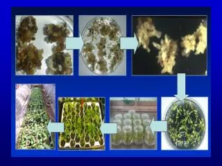
Initiation of culture Tissue dispersion Primary cell culture Subculture Cell line Continuous cell line Stored Stored Finite numbers Indefinite numbers Experimental Methods 2006
Cell Culture Morphology • Morphologically cell cultures take one of two forms: • growing in suspension (as single cells or small free-floating clumps) • cell lines derived from blood (leukaemia, lymphoma) • growing as a monolayer that is attached to the tissue culture flask. • cells derived from solid tissue (lungs, kidney), endothelial, epithelial, neuronal, fibroblasts Hela-Epithelial BAE1-Endothelial Experimental Methods 2006 SHSY5Y-Neuronal MRC5-Fibroblast
Special types of Cell culture Cells in the culture can be grown to adopt in vivo characteristic • Histotypic culture • Single cell lineage • Organotypic culture • Multiple cell lineages Experimental Methods 2006
Biology of Culture cells • Cell growth and differentiation in the culture depends on: • The nature of cells • The culture environment • the nature of the substrate on which cell grow • the physicochemical and physiological constitution of culture medium • the constitution of gas phase • the incubation temperature • the cell-cell and cell-matrix interaction Experimental Methods 2006
Cell cycle • G2 check point • DNA replicated • cell big • environment suitable • Metaphase check point • chromosome align on spindle M Mitosis G2 Gap2 G1 Gap1 G0 S Synthesis • G1 check point • cell big • environment suitable Experimental Methods 2006
Cell cycle • Interphase: • generally lasts at least 12 to 24 hours in mammalian tissue • the cell is constantly synthesizing RNA, producing protein and growing in size • Gap 0 (G0): cell will leave the cycle and quit dividing temporary or more permanent • Gap 1 (G1): Cells increase in size, RNA and protein synthesis, there is a G1 Checkpoint • S Phase: The DNA replication occurs • Gap 2 (G2): The cell will continue to grow and produce new proteins. There is a G2 Checkpoint • Mitosis or M Phase: • Cell growth and protein production stop • the cell cycle divides into two similar daughter cell • Mitosis last perhaps only one to two hours • there is a Checkpoint in the middle of mitosis (Metaphase Checkpoint) that ensures the cell is ready to complete cell division. Experimental Methods 2006
Factors affecting cell proliferation • Promotion of cell proliferation • low cell density (leaves the cell with free edge) • signals from environment: Growth factors • Inhibition of cell proliferation • Density limitation: high cell density • Contact inhibition: cell contact • signals from environment: p53 gene product Experimental Methods 2006
Factors affecting cell diferentiation • Cell differentiation is important for normal cell functions • Factors promoting cell differentiations • high cell density • cell-cell and cell-matrix interaction • inducers: hydrocortisone, retinoid, matrix Experimental Methods 2006
Factors affecting cell adhesion • Cell adhesion is important for cell proliferation and differentiation (signaling through cytoskeleton) • Cell adhesion molecule • Cell-cell interaction: CAMs, cadherins • Cell-matrix interaction: integrin, transmembrame proteoglycan • Tight junctional complex in epithelial cells for cell-cell interaction Experimental Methods 2006
Factors affecting cell adhesion • Enzymatic disaggregation digests the adhesion molecule and extracellular matrix • Most cells from solid tissues grow as adherent monolayer • Matrix-coated surface promotes cell proliferation and differentiation Experimental Methods 2006
Factors affect cell culture success • Appropriate cells • Suitable environment • Solid phase • substrate or phase on which the cell grow eg. glass, plastic, collagen, agar • Liquid phase • physicochemical and physiological constitution of the medium • Gaseous phase • Temperature • Aseptic environment Experimental Methods 2006
Solid phase • Anchorage dependent cells require a nontoxic, biologically inert to attach and allow movement for growth • The most convenient vessels are polystyrene plastic • other growth surface such as glass, filter wells • The surface can be treated by • coated with matrix substrate eg. Collagen, poly-l-lysine, matrigel • Feeder layers: monolayer of supporting cells, perhaps promote cell growth and differentiation by cell contact and substance secreted • Neurons on glial cell feeder layers Experimental Methods 2006
Liquid phase • Components of culture media • Inorganic Salts • retain the osmotic balance of the cells • regulate membrane potential by provision of sodium, potassium and calcium ions. • are required in the cell matrix for cell attachment and as enzyme cofactors. • Carbohydrates • Most media contain 4-20 mM glucose • main source of energy from glycolysis Experimental Methods 2006
Liquid phase • Proteins and Peptides • are used to replace those normally present in serum eg. transferrin, fibronectin • Amino acids • important for cell proliferation and differentiation • glutamine can enter Kreb’s cycle • Fatty Acids and Lipids • important in serum free media e.g. cholesterol and steroids essential for specialized cells. Experimental Methods 2006
Liquid phase • Vitamins • vitamins B are necessary for cell growth and proliferation • precursors for numerous co-factors • The vitamins commonly used in media include thiamine, riboflavin and biotin • Trace Elements • zinc, copper, selenium and tricarboxylic acid intermediates. • Selenium is a detoxifier and helps remove oxygen free radicals. Experimental Methods 2006
Liquid phase • Buffering Systems • most cells need optimal pH conditions in the range 7.2 - 7.4 • close control of pH is essential for optimum culture conditions • bicarbonate/CO2 buffering systems • Chemical buffering: HEPES • Most commercial culture media include phenol red as a pH indicator • yellow (acid) or purple (alkali) • Osmolarity • similar to plasma osmolarity 290 mOsm Experimental Methods 2006
Liquid phase • Serum • Undefined factors: complex mix of albumins, growth factors and growth inhibitors • increase the buffering capacity of cultures • able to bind and neutralize toxins • can be important for slow growing cells or where the seeding density is low • Subject to batch to batch variation • Heat inactivation of serum (incubation at 56ºC for 30 minutes) can help to reduce the risk of contamination Experimental Methods 2006
Gaseous phase • Carbondioxide • important for buffering system • 5-10% CO2 • Endogenous production: pyruvate • Oxygen • most cells in culture require low oxygen tension • anaerobic glycolysis • high oxygen can produce toxic free radical Experimental Methods 2006
Temperature • The optimum temperature depends on • the body temperature of animals from which the cells were obtained • anatomical variation of temperature (skin temperature may be lower than the rest of the body) Experimental Methods 2006
Aseptic techniques • Microorganism remains a major problem in cell culture • prevention of contamination • Antibiotics • improvement of laboratory condition • Aseptic techniques • Clean and tidy work surface • Personal hygiene • hand washing • caps, gowns, face mask • Reagents and media • Culture vessels Experimental Methods 2006
Cryopreservation of Cell Lines • The aim of cryopreservation is to enable stocks of cells to be stored to prevent the need to have all cell lines in culture at all times • Reduced risk of microbial contamination • Reduced risk of cross contamination with other cell lines • Reduced risk of genetic drift and morphological changes • Work conducted using cells at a consistent passage number • Reduced costs (consumables and staff time) Experimental Methods 2006
Risk Assessment Risks depend on: • Source of material • the nature of operation being carried out Assesment: • Pathogenicity • Route of transmission • Agent stability • Infectious dose • Concentration • Availability of data from animal studies • Availability of an effective prophylaxis • Medical surveillance • Experience and skill level of at-risk personnel
Risk groups for animal cell culture • The level of risk depends on the cell line to be used and is based on whether the cell line is likely to cause harm to humans. • Low risk • Non human/non primate continuous cell lines and some well characterized human diploid lines of finite lifespan • Medium risk • Poorly characterized mammalian cell lines. • High risk • Cell lines derived from human/primate tissue or blood. • Cell lines with endogenous pathogens (the precise categorization is dependent upon the pathogen) • Cell lines used following experimental infection where the categorization is dependent upon the infecting agent Experimental Methods 2006
Safety aspects of cell culture • SAFETY CONSIDERATIONS • Assume all cultures are hazardous since they may harbor latent viruses or other organisms • The following safety precautions should also be observed: • pipetting: use pipette aids to prevent ingestion • keep aerosols down to a minimum • no eating, drinking, or smoking • wash hands after handling cultures and before leaving the lab • decontaminate work surfaces with disinfectant (before and after) • autoclave all waste • use biological safety cabinet (laminar flow hood) • use aseptic technique • dispose of all liquid waste after each experiment and treat with bleach Experimental Methods 2006
Risk Group (RG) Classification is based on the potential effect of biological agent on healthy human adult • RG1-agents are not associated with disease • RG2-agents are associated with human disease which is rarely serious and for which preventive or therapeutic interventions are often available • RG3-agents are associated with serious or lethal human disease for which preventive or therapeutic interventions may be available • RG4-agents are likely to cause serious or lethal human disease for which preventive or therapeutic interventions are not usually available
Biosafety cabinets • The Class I BSC provides personnel and environmental protection, but no product protection. • The Class I BSC is hard-ducted to the building exhaust system, thimble-connected, or recirculated back into the room depending on use. • The Class II (Types A, B1, B2, and B3)24 biological safety cabinets provide personnel, environmental and product protection. Laminar flow
BSL Agents Practices Safety Equipment(Primary Barriers) Facilities (Secondary Barriers) 1 Not known to consistently cause disease in healthy adults Standard Microbiological Practices None required Open bench top sink required 2 Associated with human disease, hazard = percutaneous injury, ingestion, mucous membrane exposure BSL-1 practice plus:• Limited access • Biohazard warning signs• "Sharps" precautions • Biosafety manual defining any needed waste decontamination or medical surveillance policies Primary barriers = Class I or II BSCs or other physical containment devices used for all manipulations of agents that cause splashes or aerosols of infectious materials; PPEs: laboratory coats; gloves; face protection as needed BSL-1 plus: Autoclave available 3 Indigenous or exotic agents with potential for aerosol transmission; disease may have serious or lethal consequences BSL-2 practice plus:• Controlled access• Decontamination of all waste• Decontamination of lab clothing before laundering• Baseline serum Primary barriers = Class I or II BCSs or other physical containment devices used for all open manipulations of agents; PPEs: protective lab clothing; gloves; respiratory protection as needed BSL-2 plus:• Physical separation from access corridors• Self-closing, double-door access• Exhausted air not recirculated• Negative airflow into laboratory 4 Dangerous/exotic agents which pose high risk of life-threatening disease, aerosol-transmitted lab infections; or related agents with unknown risk of transmission BSL-3 practices plus:• Clothing change before entering• Shower on exit• All material decontaminated on exit from facility Primary barriers = All procedures conducted in Class III BSCs or Class I or II BSCs in combination with full-body, air-supplied, positive pressure personnel suit BSL-3 plus:• Separate building or isolated zone• Dedicated supply and exhaust, vacuum, and decon systems• Other requirements outlined in the text Recommended Biosafety Levels for Infectious Agents
References • R. Ian Freshney. Culture of Animal cells a manual of basic technique. 4th edition. Wiley-Liss, New York. 2000. Experimental Methods 2006
Tissue culture Experimental Methods 2006
P2 Room Experimental Methods 2006