Slime molds
Slime molds. Slime molds. Five phyla of organisms (we will discuss two) that are not in the Kingdom Fungi, but Kingdom Protoctista Vegetative thallus – lacks a cell wall, amoeba like, phagotrophic, i.e. ingests food particles by phagocytosis May be multinucleate = plasmodium

Slime molds
E N D
Presentation Transcript
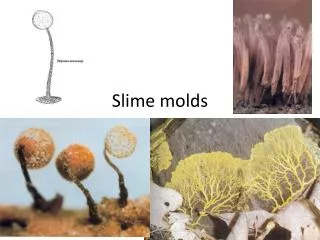
Slime molds • Five phyla of organisms (we will discuss two) that are not in the Kingdom Fungi, but Kingdom Protoctista • Vegetative thallus – • lacks a cell wall, • amoeba like, • phagotrophic, i.e. ingests food particles by phagocytosis • May be multinucleate = plasmodium • Have primarily been studied by mycologists, found in habitats of fungi, some produce fruiting structures that resemble fungi
Slime mold phyla • Dictyosteliomycota – cellular slime molds • Vegetative thallus – amoebae that aggregate to form pseudoplasmodium • 3 genera, 50 spp. • Myxomycota – true slime molds • Vegetative thallus – plasmodium • 71 genera, 500 spp.
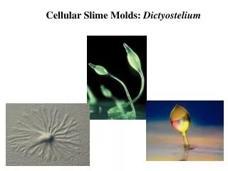
Dictyosteliomycota – cellular slime molds • Widely used in studies of eukaryotic cell development • Make transition from population of individual amoeboid cells to multicellular structure • Occurrence – widespread in forest soils, dung, decaying plant matter • Feed on bacteria in soil as amoeboid cells
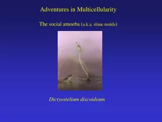
Dictyostelium discoideum life cycle • Vegetative thallus – unicellular amoebae that feed on bacteria by phagocytosis • Asexual reproduction – cell division • Can form microcysts – form thin cellulose cell wall & withstand unfavorable environmental conditions
Developmental changes • When food supply becomes exhausted or population reaches certain size, amoebae enter a starvation period • Amoebae undergo developmental changes • Metabolic changes – shift from facultative aerobes to obligate aerobes • Use endogenous reserves • Cell surface antigens change – cells become more cohesive • Certain amoebae secrete a chemotactic substance - acrasin
Aggregation • Acrasin in Dictyostelium is cyclic AMP, other species produce other substances • Acrasin causes other amoebae to migrate toward the center of production in pulsating streams – aggregation stage
Pseudoplasmodium • Amoebae aggregate to form pseudoplasmodium (slug, grex) • Transition from population of independent cells to a multicellular structure • Pseudoplasmodium in D. discoideum is 1-2 mm long and moves along gradients of temperature, light, humidity • Is surrounded by a sheath of polysaccharide and protein, • Leaves a trail of slime as it migrates
Pseudoplasmodium • Amoebae do not feed or divide • If food is added, may be de-aggregated up to a certain point after which they are committed to development • As the slug migrates, it becomes polarized and cells begin to differentiate
Differentiation • Two cytologically and biochemically distinct types of cells are forming in slug • Prestalk cells – anterior portion (1/3) of slug • Swell, form a cell wall, become vacuolate and eventially die as they become stalk cells • Prespore cells – posterior portion (2/3) of slug • Form prespore vacuoles – involved in cell wall synthesis
Culmination • Slug migration ceases and becomes globose • Prestalk cells form the beginning of the stalk
Sorocarp • Stalk cells are formed, prespore cells migrate up the stalk • Prespore cells form cellulose cell wall become spores • Structure formed is a sorocarp with spores in the sorus (droplet containing spores) – not enclosed by wall, not a sporangium • Spores • are uninucleate • remain dormant • Germinate to form an amoeba
Sorocarp • Formation of sorocarp – for dispersal of spores • Asexual reproduction occurs as a result of cell division by amoebae before sorocarp formation • Ca. one third of amoebae lost in sorocarp formation (produce stalk) • Stalk is cellular
Sexual reproduction • Not well understood • Giant cells (zygotes) formed from fusion of two amoebae (gametes) • Large number of amoebae migrate to zygote, secrete wall to enclose amoebae and zygote • Zygote feeds on amoebae • Other wall layers produced to form macrocyst
Macrocyst • Meiosis occurs in macrocyst • Cytoplasm cleaves to produce uninucleate amoebae • Amoebae released through broken cyst walls • Both homothallic and heterothallic strains are known from different species
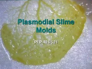
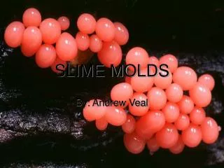
Myxomycota – true slime molds • Produce a true plasmodium at some point in their life cycle • Plasmodium – multinucleate mass of protoplasm that feeds by phagocytosis • Great variability in size – some are microscopic, others may grow to meters
Habitats • Commonly occur in cool, moist shady habitats, e.g. decaying logs • Can occur on lawns if weather is moist • Have also been found on bark of trees and in deserts • Feed on bacteria, protozoa, small pieces of organic matter • Generally not of great economic importance
Life cycle • Two amoeba-like vegetative phases • Plasmodium • Myxamoebae • Complex fruiting structures – sporophores • Few species have been cultured (dual cultures) and grown through all stages in life cycle • Fewer have been grown in axenic culture
Spores • Spores are haploid, spherical • Thick walled with spines, teeth, other ornamentation • Cell wall composition not well known – one report – galactosamine polymer & melanin • Can remain dormant (to at least 75 yrs)
Spore germination • Cell wall either splits or small pore is digested • Germination produces amoeba-like cells - myxamoebae (one or several) – or swarm cells – that have 2 flagella • Myxamoebae and swarm cells can be interconverted – when water present, flagella are produced
Myxamoebae • Feed by phagocytosis of bacteria, other small particles • Divide by mitosis – dissolution of nuclear membrane and formation of centrioles • If unfavorable conditions occur – can encyst (form a cell wall) to form a microcyst
Sexual reproduction • Plasmogamy occurs between myxamoebae or swarm cells (some species are heterothallic) • Must also be a critical mass of cells in population • Karyogamy occurs shortly after plasmogamy to form zygote (2n) • Zygote feeds, can engulf other myxamoebae, coalesce with other zygotes
Plasmodium • Zygote forms plasmodium – longest lived vegetative stage • Variation in species – • Size – microscopic to meter across • Color – colorless, black, violet, red, yellow, etc • No definite shape • Move over surface engulfing particles • Vein like network with viscosity differences in cytoplasm • Rapid cytoplasmic streaming
Plasmodium • Phagocytosis of particles • Can absorb nutrients • Nuclei divide in synchronous fashion • In mitosis, nuclear envelope doesn’t break down, no centrioles
Sclerotium • Unfavorable environmental conditions can induce plasmodia to form dormant structures - sclerotia • Hardened mass containing spherules – cytoplasm and several nuclei surrounded by cell wall • Favorable conditions – germinate to form plasmodia
Sporulation • Entire plasmodium differentiates to form reproductive structures • Environmental conditions trigger – moisture, light, temperature, pH, exhaustion of food supply • Sporulating structures = sporophores, 3 types • Sporangium (pl. sporangia) • Aethallium (pl. aethallia) • Plasmodiocarp
Sporophores • In all sporophores, the multinucleate cytoplasm is cleaved into many spores • Membranes are laid down around nuclei • Cell walls are formed around cell membrane • This differentiates a sporangium from a sorus
Sporangia • Most common type of sporophore • One plasmodium may form many sporangia • Parts of a sporangium • Hypothallus – secretion of plasmodium that is left on substratum, base of the sporangium, may be a thin, cellophane-like secretion or a crust of CaCO3
Parts of a sporangium • Stalk – supports sporangium, may or may not be present, may be hollow or filled with material • Stalks formed from secretions of plasmodium and are acellular (in contrast to cellular slime molds)
Parts of a sporangium • Peridium – outer covering of sporangium • Ranges from delicate membrane to tough covering • May be impregnated with CaCO3
Parts of a sporangium • Columella – an extension of the stalk into the sporangium
Parts of a sporangium • Capillitium – nonliving threads that intermingle but are not attached to spores • May be attached to peridium or columella • May be ornamented • Formed during spore cleavage • Involved in dispersal
Parts of a sporangium • Spores – main function of sporophore is to form spores • Formed from a multinucleate mass of cytoplasm – vacuoles form and fuse to form membranes around nuclei, cell walls formed in vacuoles – called cleavage
Spores • Are uninucleate and diploid at first • Meiosis occurs to form 4 haploid nuclei • Three nuclei may disintegrate to form uninucleate haploid spores • In some, spore may contain more than one nucleus – germinate to produce more than 1 myxamoebae
Other types of sporophores • Aethallium – fairly large cushion shaped structure (does not differentiate into individual sporangia)
Other types of sporophores • Plasmodiocarp – similar in appearance to plasmodium, plasmodial veination is retained, stalkless
Classification • Myxomycota classified on the characteristics of their sporophores – presence or absence of capilltium, stalk, nature of the peridium, etc. • Many form brightly colored sporophores