Download
1 / 14
150 likes | 337 Views
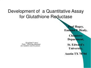
Development of a Quantitative Assay for Glutathione Reductase. Paul Hagey, Eamonn F. Healy, Chemistry Department, St. Edward’s University, Austin TX 78704. Abstract.

E N D
Development of a Quantitative Assay for Glutathione Reductase Paul Hagey, Eamonn F. Healy, Chemistry Department, St. Edward’s University, Austin TX 78704
Abstract Glutathione Reductase has been implicated as the critical enzyme for maintenance of GSH during oxidative stress. We report here steps towards development of quantitative assay for Glutathione Reductase using biotin. Biotinylation has achieved widespread and general utility in a variety of bioanalytical applications due to the ready formation of biotin-avidin complexes (1015 M-1). We have succeeded in coupling a biotin to GSSG (reduced glutathione), with detection by HPLC at 260nm. The GSSG-biotin is then complexed with Glutathione Reductase, and subsequently complexed in a Glutathione Reductase :GSSG-biotin : avidin triplex. By purifying the resulting complex and spectroscopically determining the concentration, Glutathione Reductase can be accurately assayed.
Glutathione Reductase Glutathione reductase (GR, EC 1.6.4.2) is an ubiquitous enzyme which catalyzes the reduction of oxidized glutathione (GSSG) to glutathione (GSH). Glutathione reductase is essential for the glutathione redox cycle that maintains adequate levels of reduced cellular GSH. GSH serves as an antioxidant, reacting with free radicals and organic peroxides, in amino acid transport, and as a substrate for the glutathione peroxidases and glutathione S-transferases in the detoxification of organic peroxides and metabolism of xenobiotics, respectively
Biotinylated Probes • Biotin (B) is a small molecule that covalently attaches to selected residues of bioactive peptides and proteins , termed probes (P), without causing loss of function • Biotin is useful because its high affinity for avidin (Av) and streptavidin allows the biotin-peptide complex to form a triplex (Av:B-P) with the protein, thus isolating the probe • By incubating this triplex with the probe’s natural target molecule (T) a complex of composition Av:B-P:T is now isolated • An assay of the avidin present is now also a quantitative assay the target protein
Project • Our first requirement was to identify the particular form of biotin (B) suitable for attachment to our GSSG peptide. We have achieved success with a NHS-ester derivative with a 6-C spacer arm, shown on the right. • After developing a suitable HPLC separation method we are currently focusing on using preparative HPLC to isolate the purified B-GSSG probe. • After suitable characterization we will proceed to form a triplex (Av:B-GSSG) of a out biotinylated probe with Avidin • Finally we hope to form our GRase:GSSG-B:Av complex
HPLC HPLC instrumentation includes a pump, injector, column, detector and recorder or data system, connected as shownon the right. The heart of the system is the column where separation occurs. The chromatographic process begins by injecting the solute onto the top of the column.Separation of components occurs as the analytes and mobile phase are pumped through the column. Eventually, each component elutes from the column as a narrow band (or peak) on the recorder. Analyte molecules, while moving through the porous packing bead, tend to interact with the surface adsorption sites. All these interactions are competitive. Analyte molecules are competing with the eluent molecules for the adsorption sites. So, the stronger analyte molecules interact with the surface, and the weaker the eluent interaction, the longer analyte will be retained on the surface.
Biotinylation Methodology • The probe to be biotinylated is dissolved in a phosphate, pH=7.6 buffer at a concentration of 10mg/mL • The biotinylation reagent is dissolved in dimethylformamide at a concentration of 25mg/mL, and stored as a stock solution at 2-8 oC (stable for approx. 24 hrs) • The biotinylation reagent is added slowly to the probe solution in a 10-30 molar excess and gently mixed for 1-4 hours • The reaction is terminated by adding 1mL of 20% TFA • The products are analyzed using reverse phase HPLC and a C-18 column
Results GSSG Time :1 hour Phosphate pH=7.6 Solvent A: ACN Solvent B: ACN/1%TFA Detector : 260nm Flow Rate : 1ml / min Biotin Time : 1hour Phosphate pH=7.6 Solvent A: ACN Solvent B: ACN/1%TFA Detector : 260nm Flow Rate : 1ml / min
Results GSSG / Biotin Time : 1 day Phosphate pH=7.6 Solvent A: ACN Solvent B: ACN/1%TFA Detector : 260nm Flow Rate : 1ml / min GSSG / Biotin Time : 4 days Phosphate pH=7.6 Solvent A: ACN Solvent B: ACN/1%TFA Detector : 260nm Flow Rate : 1ml / min
Results Lys-Tyr-Lys / Biotin Time :initial Phosphate pH=7.6 Solvent A: ACN Solvent B: ACN/1%TFA Detector : 260nm Flow Rate : 1ml / min Lys-Tyr-Lys / Biotin Time : 1 day Phosphate pH=7.6 Solvent A: ACN Solvent B: ACN/1%TFA Detector : 260nm Flow Rate : 1ml / min
Discussion • The peak at 7.6 min on the chromatogram of the GSSG / NHS-spacer-biotin indicates to use the successful biotinylation of our peptide probe. However definitive characterization will require purification using a semi-preparative C-18 column and analysis by mass spectroscopy. • As a preliminary check we biotinylated a Lys-Tyr-Lys tripeptide, similar in size to GSH, and found a peak at a retention time of 8.1 mins. Since biotin is known to attach easily to the e-amino group of a lysine residue this result does seem to support our hopes of a successful biotinylation of GSSG. It remains to bee seen whether this 7.6 min peak represents a mono-or di-biotinylated adduct. • Upon purification of the B-GSSG complex we intend to incubate it with avidin horseradish peroxidase(AvHpr), an avidin derivative capable of simple and quantitative assay • Final formation of our Grase:GSSG-B:AvHpr complex will then allow a quick, colorimetric, quantitive assay of Glutathione Reductase
References • Brian T. Miller, et al.;Peptide Biotinylation with Amine-Reactive Esters;Peptides. 1997, 18, 1586 Acknowledgements • We gratefully acknowledge the support of the Welch Foundation in the form of a Departmental Research Grant