Download
1 / 9
90 likes | 249 Views
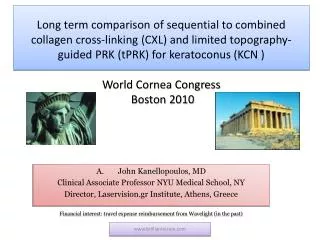
Novel Cornea OCT (cOCT) findings in early and long term follow up of collagen cross-linking (CXL) for keratoconus (KCN) ASCRS, Boston 2010. John Kanellopoulos, MD Associate Professor of Ophthalmology, NYU Medical School, NY, NY, USA Director, Laservision.gr Institute, Athens, Greece.

E N D
Novel Cornea OCT (cOCT) findings in early and long term follow up of collagen cross-linking (CXL) for keratoconus (KCN)ASCRS, Boston 2010 John Kanellopoulos, MD Associate Professor of Ophthalmology, NYU Medical School, NY, NY, USA Director, Laservision.gr Institute, Athens, Greece Financial interest: travel expense reimbursement from Wavelight(in the past) Kanellopoulos MD www.brilliantvision.con
Introduction • The depth and degree of collagen cross-linking has been difficult to evaluate in clinical reality. By scanning some of these corneas with cOCT we encountered different findings than non-cxled corneas. We are therefore herein attemping to evaluate these novel, reproducible cornea OCT findings the first 6 months and at least 3 years following CXL (collagen cross-linking) for keratoconus Kanellopoulos MD www.brilliantvision.con
Methods We evaluated a total of 125 KCN cases frm our clinical practice; 70 recent (group A) and 55 treated earlier (group B) were evaluated for the presence and stromal depth of intrastromal CXL lines (I-CXL-l), cornea thickness in microns (CT), UCVA, BSCVA, keratometry (K), topography, endothelium (ECC) and clarity. The Optovue OCT devise was used in this study Kanellopoulos MD www.brilliantvision.con
Results 68 of Group A and 51 of group B cases demonstrated I-CXL-l. The mean values for group and group B were respectively: CT : 370, 380, I-CXL-l depth: 255, 262, UCVA: 20/60, 20/50, BSCVA: 20/40, 20/30, K: 49.5, 48.5, ECC: 2550, 2600. Mean follow-up: 7 , 38 months respectively Kanellopoulos MD www.brilliantvision.con
A cornea OCT demonstrating hyper-reflective intra-corneal stromal “lines” at 2/3 depth (yellow arrows) corresponding with the clinical presence of CXL demarcation line in a patient, 3 years following the combined Topography guided-PRK/ and CXL procedure. Kanellopoulos MD www.brilliantvision.con
Other examples Kanellopoulos MD www.brilliantvision.con
Conclusions cOCT appears to demonstrate reproducible early and long-term CXL cornea findings. The hyper-reflective “lines’ may represent induced cornea density or subtle intrastromal cornea scarring This may constitute a possible novel non-invasive measurement, to evaluate and titrate the amount, extent and depth of intra-stromal effects of the CXL treatment in keratoconic and possibly ectasia eyes. Kanellopoulos MD www.brilliantvision.con
Thank you www.brilliantvision.com Kanellopoulos MD www.brilliantvision.con