Download
1 / 30
320 likes | 994 Views
QUESTION 1. Identify the radiopaque entity. Genial tubercles Lingual foramen Mental foramen Mylohyoid ridge. QUESTION 2. Identify the radiopaque entity. Condyle Coronoid process Maxillary tuberosity Mandibular molar . QUESTION 3. Identify the radiopaque entity. Mylohyoid ridge

E N D
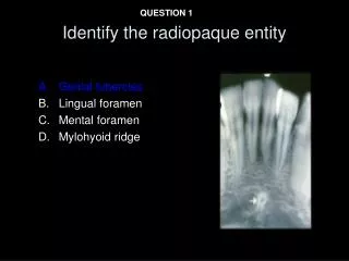
QUESTION 1 Identify the radiopaque entity • Genial tubercles • Lingual foramen • Mental foramen • Mylohyoid ridge
QUESTION 2 Identify the radiopaque entity • Condyle • Coronoid process • Maxillary tuberosity • Mandibular molar
QUESTION 3 Identify the radiopaque entity • Mylohyoid ridge • External oblique ridge • Submandibular salivary gland fossa • Genial tubercles
QUESTION 4 Identify the radiolucent space • Maxillary sinus • Lateral border of the nose • Zygomatic process of the maxilla • Posterolateral wall of the maxillary sinus
QUESTION 5 Identify the radiopaque entity • Floor of the maxillary sinus • Lateral border of the nose • Zygomatic process of the maxilla • Posterolateral wall of the maxillary sinus
QUESTION 6 Identify the abnormality • Associated teeth are vital • A.Rarefying osteitis • B. Osteosclerosis • C. Periapical cemento-osseous dysplasia • D. Osteomyelitis
QUESTION 7 Identify the radiopaque entities • A. Periodontal ligament space • B. Remnants of Lamina dura • C. Bone resorption
QUESTION 8 Localize the radiopaque entity • Buccal • Lingual
QUESTION 9 Identify the radiolucent area • Inferior alveolar nerve canal • Submandibular gland • Submandibular salivary gland fossa • External oblique ridge
QUESTION 10 Identify the abnormality • A. Cherubism • B. Treacher- Collin syndrome • C. Basal cell nevus syndrome
QUESTION 11 Identify the radiolucent line • Intermaxillary suture • Nasopalatine duct • Incisive foramen The difference between suture and canal: The suture is an area where two bones come together. It is a radiolucent line And that line can be thin or thick for example Both these lines can represent a suture. On the other hand a canal would be like a tunnel within bone. It will have bone all around it thus there will be two radiopaque lines and a RELATIVE radiolucency within (since there is buccal and lingual bone around it). A canal is hollow tube within bone. Also the nasopalatine canal starts approx. from the level of the middle 3rd of the roots of the central incisors.
QUESTION 12 Identify the radiopaque entity • Inferior nasal conchae • Nasal septum • Middle nasal conchae
QUESTION 13 Identify the radiopaque border • Floor of the maxillary sinus • Lateral border of the nose • Zygomatic process of the maxilla • Posterolateral wall of the maxillary sinus
QUESTION 14 Identify the abnormality • Associated tooth has no decay or fracture. • A.Rarefying osteitis • B. Sclerosing osteitis • C. Osteosclerosis • D. Osteomyelitis
QUESTION 15 Identify the abnormality • 35 year old female. Associated teeth are vital • A.Rarefying osteitis • B. Osteosclerosis • C. Periapical cemento-osseous dysplasia • D. Osteomyelitis
QUESTION 16 Identify the abnormality A. Compound odontoma B. Osteosclerosis C. Sclerosing osteitis D. Complex odontoma
QUESTION 17 Identify the abnormality A.Enostosis B. Osteosclerosis C. Exostosis D. Sclerosing osteitis
QUESTION 18 Identify the abnormality 1. Central giant cell lesion 2. Odontogenic myxoma 3. Odontogenic keratocyst 4. Ameloblastoma A. 1, 2 &3 B. 1, 2, 3 &4 C. 1 & 2 only D. 3 &4 only
QUESTION 19 Identify the abnormality A.Osteosclerosis B. Cementoblastoma C. Sclerosing osteitis D. Enostosis
QUESTION 20 Identify the entity • Ascending ramus of mandible • Condyles • Coronoid process • Body of the mandible
QUESTION 21 Identify the abnormality A.Normal trabecular bone B. Fibrous dysplasia C. Sclerosing osteitis D. Osteosclerosis
QUESTION 22 Identify the abnormality A.Hypercementosis B. Cementoblastoma C. Sclerosing osteitis D. Enostosis
QUESTION 23 Identify the abnormality 1. Central giant cell lesion 2. Odontogenic myxoma 3. Odontogenic keratocyst 4. Ameloblastoma A. 1, 2 &3 B. 2, 3, 4 &1 C. 1 & 2 only D. 3 &4 only
QUESTION 24 Identify the entity • Identify the radiopaque entity • Inferior nasal conchae • Nasal septum • Middle nasal conchae
QUESTION 25 Identify the process • (Associated soft tissue mass in oral cavity) • Benign • Malignant
QUESTION 26 Identify the abnormality • Gardner’s syndrome • Neurofibromatosis • Paget disease of bone
QUESTION 27 Identify the abnormality • Patient has high alkaline phosphatase levels and hypercementosis on multiple teeth • Fibrous dysplasia • Paget disease of bone • Multiple myeloma
QUESTION 28 Identify the abnormality A.Intraosseous hemangioma B. Extraosseous hemangioma C. Enostosis D. Exostosis
QUESTION 29 Identify the abnormality • Squamous cell carcinoma • Plasmacytoma • Multiple myeloma • Mucoepidermoid carcinoma
QUESTION 30 Identify the abnormality • Squamous cell carcinoma • Leukemia • Multiple myeloma • Mucoepidermoid carcinoma