Download
1 / 64
650 likes | 835 Views
Mammography. Introduction and History. Breast cancer is 2nd only to lung cancer as cause of death in women Very treatable with early detection! 1st innovation since radical mastectomy introduction in 1898 In 1913, radiographic appearance of breast cancers was first reported

E N D
Introduction and History • Breast cancer is 2nd only to lung cancer as cause of death in women • Very treatable with early detection! • 1st innovation since radical mastectomy introduction in 1898 • In 1913, radiographic appearance of breast cancers was first reported • Mammography became a reliable diagnostic tool in 1950s when industrial grade x-ray film introduced
History Of Mammography (cont’d) • 1960’s – Xerography introduced – much lower dose • Research conducted in 1970s clearly showed mammography to be essential part of early diagnosis • 1975 – High speed/resolution film introduced by DuPont • 1992 – MQSA implemented (MammographyQuality Standards Act)
Definition of breast cancer: • Cancer that forms in tissues of breast, usually ducts (tubes that carry milk to nipple) and lobules (glands that make milk). • Occurs in both men and women(male breast cancer is rare)
MQSA • Mammography was 1st and only federally regulated imaging exam with implementation of Mammography Quality Standards Act(MQSA) • Mandated following: • Formal training and continuing education • Required regular inspection of equipment • Documentation of quality assurance • Reporting results, follow-up, tracking pts, and monitoring outcomes
Principles Of Breast Cancer • Pt.s inearly stagesrespondwellto treatment • Patients withadvanced diseasedopoorly • Earlier diagnosis, better chance of survival • Mammographyis tool for early detection
Risk v. Benefit • Breast cancer in United States in 2009 (estimated): New cases: 192,370 (female); 1,910 (male) Deaths: 40,170 (female); 440 (male) • Us population 306 million in 2007-133 deaths /million • Mortality risk from mammography induced radiation is 5 deaths/ million pts. using screen film mammography • More risky to refusemammography!
Breast Cancer Screening • Very 1st Mammogram is Baseline (or first mammo. after surgery) • There after: screening mammogram pt. must be asymptomatic – no known breast problems • American Cancer Society and American College of Radiology recommend screening annually for women over age 40
Diagnostic Mammogram • For woman presenting with clinical evidence ofbreast disease, palpable mass or other symptom • Uses specific projections to • Rule out cancer • Demonstrate suspicious area seen on screening mammogram
Breast Cancer Risk Factors • Risk increases with age • Hormonal history • Risk increases with early menses, late menopause, pregnancy after age 30, or nulliparity • Family history • Risk increases -daughter, mother, or sister has breast cancer
Breast Anatomy • Breast same as mammary gland • Lobulated, glandular structures located in superficial fascia of anterolateral wall of thorax • Secondary sex characteristic
Base of breast overlies pectoralis major and serratus anterior muscles • Part of breast extends into axillary fossa
Anatomy (cont’d) • Breasts vary in size and shape! • Consist of glandular, fat, and muscle tissue
Breast Anatomy • Lobule size affected by age and hormones • Involution: process of decreasing lobule size with age and after pregnancy
Anatomy • The breast tapers anteriorly ending in the nipple • Encircled by areola: area of pigmented skin • Breasts are supported by Cooper’s ligament which determines firmness or lack thereof • Female breasts are divided into 15 – 20 lobules
Breast Anatomy • Each lobe divided into many lobules • Lobules are basic structural unit of breast • Lobules contain • Several acini • Draining ducts • Interlobule stroma (connective tissue)
Breast Anatomy • Lymphatic vessels of breast drain into two sets of nodes • Axillary lymph nodes, laterally • Internal mammary lymph nodes, medially • Axillary nodes are often evaluated on mammograms
Tissue Variations • Breasts -glandular and connective • Ability to visualize depends upon amount of fat within and around breast lobules- provides contrast • Postpuberty breasts contain primarily dense connective tissue
Fatty tissue replaces glandular tissue after lactation and advancing age • After menopause, glandular tissue begins to atrophy
Typical Mammography Unit Equipment is C-arm SID is fixed at 24 – 26”
Mammography Equipment • Dedicated units have high-frequency generators • Provide more precise control of kVp, mA, and exposure time • Specially designed to produce high-contrast and high-resolution images
Mammography uses • Low kVp : 25 – 28 • AEC • Anode material made of molybdenum, with rhodium target • Grid with ratio: 4:1, or 5:1 200 lines/inch
Magnification • Increases visibility of small structures • Increase OID • Uses air gap • Radiation dose increases with magnification
Compression Device • Compression decreases thickness of breast, magnification and scatter • Increases contrast • Reduces motion unsharpness • Reduces dosage
Compression Device Made of firm plastic Amount of compression: between 25 and 40 pounds pressure Compression may beuncomfortable!
Screen-Film Systems • Mammography cassettes contain a single screen • Film is single emulsion • Occasionally, extended time processing is used • (reduces dose and increases contrast)
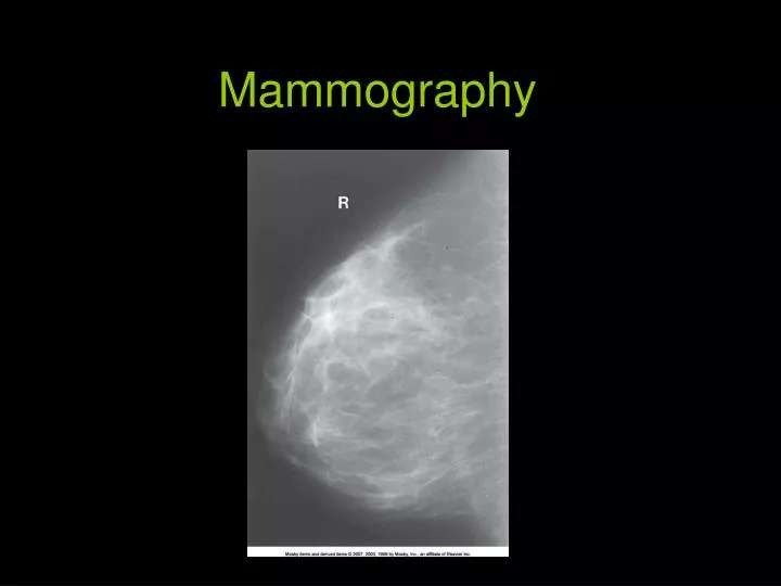
Digital Mammography State of the art! • No film or chemical processing • Images easily sent over internet • Much better definition Possible downside-if 1st digital compared to previous film mammo., can give false positives due to increased sensitivity! - Slightly higher dosage
Procedure • Complete, careful history and physical assessment • Take notes on location of scars, palpable masses, skin abnormalities, and nipple alterations • Examine previous mammograms for positioning, compression, and exposure factors
Procedure (con’t) • Patients dress in open-front gown • Breasts must be bared for imaging • Cloth will cause image artifact • Remove deodorant and powder from axilla and breast • Can mimic calcifications on image
Procedure (cont’d) • Explain procedure to pt., including possibility for additional projections • Consider natural mobility of breast before positioning • Support breast firmly so that nipple is directed forward • Profile nipple, if possible
Procedure • Apply proper compression to produce uniform breast thickness • Essential to high-quality mammograms • Place ID markers according to standard convention
Routine mammography projections Craniocaudal (CC) Mediolateral oblique (MLO)
Craniocaudal Projection Patient position • Standing or seated facing IR holder • Part position • Elevate inframammary fold to maximum height • Adjust IR height to inferior surface of breast • Gently pull breast onto IR holder with both hands while instructing patient to press chest to IR holder
Craniocaudal Projection • Arrange breast on film so nipple is in profile and maximum amount of breast tissue is radiographed • CR – Perpendicular to base of breast • Structures – Central, subareolar, medial fibroglandular breast tissue, pectoral muscle
Craniocaudal Positioning (cont’d • Immobilize breast with one hand • Use other hand to move opposite breast out of image • Shoulder relaxed in external rotation
Craniocaudal Projection (cont’d) • Rotate head away from breast being examined (watch out for hair!) • Lean pt. toward machine • Place hand on shoulder and slide skin over clavicle • Compress breast slowly until skin taut
Mediolateral Oblique Projection • Position • Center breast with nipple in profile, if possible • Hold breast up and out • Compress breast slowly until taut • Pull down on abdominal tissue to open inframammary fold
Mediolateral Oblique positioning • Instruct pt. to hold opposite breast laterally, out of anatomy of interest • Exposure on suspended respiration • Release compression immediately!
Mediolateral Oblique • Open inframammary fold • Deep and superficial breast tissues well separated • Retroglandular fat well seen • Uniform tissue exposure • If compression is adequate
Mediolateral Oblique • Degree of obliquity is 30° to 60° • Depends on body habitus • Tall, thin patients require steeper angulation • CR perpendicular to base of breast • Structures – lateral aspect of breast and axillary tail
Radiography Of Augmented Breast (implants) • 8 projections must be obtained (2x4) • MRI and sonography can help determine rupture or leakage • Four standard images with implant displaced posteriorly into chest wall are obtained
Saline vs Silicone • Some surgeons feel silicone implants have a morenatural look and feel because silicone gel texture similar to breast tissue. • Silicone implant ruptures are harder to detect. When saline implantsrupture, they deflate -results are seen almost immediately. When silicone implants rupture, breast often looks and feels same because silicone gel may leak into surrounding areas of breast without a visible difference. • Replacing a ruptured silicone gel implant is more difficult than repairing saline implant. Silicone implants have higher rate of capsular contracture (scarring and hardening around implant). • Saline implants inflated to desired size with saline, then valve is sealed by surgeon
Radiography Of Augmented Breast (implants) Complications: • Increased fibrous tissue surrounding implant (contracture) • Shrinking • Hardening • Leakage • Pain
Male Mammography • Approximately 1000 males develop breast cancer every year • Standard CC and MLO are obtained • Males not screened- mammogram only if lump discovered