Download
1 / 60
630 likes | 905 Views
Key Stage 4. The Breathing System. Teacher’s Notes. A slide contains teacher’s notes wherever this icon is displayed - To access these notes go to ‘Notes Page View’ (PowerPoint 97) or ‘Normal View’ (PowerPoint 2000). Notes Page View. Normal View. Flash Files.

E N D
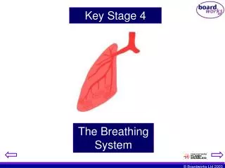
Key Stage 4 The Breathing System
Teacher’s Notes A slide contains teacher’s notes wherever this icon is displayed - To access these notes go to ‘Notes Page View’ (PowerPoint 97) or ‘Normal View’ (PowerPoint 2000). Notes Page View Normal View Flash Files A flash file has been embedded into the PowerPoint slide wherever this icon is displayed – These files are not editable.
Humans breathe to ensure that oxygen enters the body and that carbon dioxide leaves the body. Oxygen (O2) Carbon Dioxide (CO2)
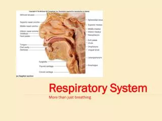
The breathing system HEAD THORAX ABDOMEN Let us now look at the structure of the breathing system. The human body can be divided into three regions. The breathing system is found in the thorax.
The body separates the process of breathing in and breathing out. Breathing in is one process and is known as… Inhalation (When we breathe in we inhale) Breathing out is another separate process and is known as… Exhalation (When we breathe out we exhale) By separating these two processes, the body can concentrate on the two tasks in turn.
Firstly it must inhaleoxygen and secondly it must exhalecarbon dioxide The breathing system is designed to be able to perform both tasks using the same organs. One final important fact to remember is that breathing can be performed without humans having to think about it. Just imagine that as well as everything else you have to think about, you would have to remember to tell your body to inhale, then exhale, then inhale, exhale, inhale, … etc. There would be no time for anything else.
So, what does this system look like? Well, let us start where air enters the system… Air enters through either the mouth or the nostrils. Nasal cavity Nostril Mouth Oral Cavity It does not matter through which opening the air enters because the oral and nasal cavities are connected.
As the air passes through the nasal cavity, the air is smelt, warmed, filtered and moistened slightly. The air meets at the Pharynx, a junction at back of the oral cavity. The Pharynx is a junction between two tubes. The air must travel down only one of these tubes. One is the Windpipe (Trachea) and the other is the Gullet (Oesophagus) Trachea Gullet As the name suggests, air must pass down through the windpipe (trachea).
You can think of the trachea as a tube lined with C-shaped supporting rungs. These rings are made of a tough material called Cartilage. They help to hold the tube open. Diagram of trachea with cartilage rungs. You may be wondering why they are C-shaped and not full circles.
Well, if a tube were lined with fixed circles of cartilage, it would have a fixed diameter… Although this would stop the tube from collapsing, this would also mean that the tube would not be able to expand. When we breathe in, the trachea must expand to allow more air in. Cartilage Trachea
Therefore, a C-shaped piece of cartilage can change shape.
As well as being adapted on its outer surface, the trachea shows adaptations on its inner lining. If we look closely at the inner surface of the trachea… ciliated epithelial cells
The cells that line the wall of the trachea show two special adaptations. tiny hairs called Cilia produce a sticky liquid called mucus Ciliated epithelial cells We say the cells show specialisation. These specialised cells have a particular job to do.
The presence of mucus and cilia on the lining of the trachea ensures that the air we breathe is clean and free fromdisease. Microbes travelling down the trachea within inhaled air. Microbes become stuck within the mucus. Ciliated Cells Mucus being made by the ciliated cells.
Once the microbes are stuck in the mucus, the cilia move the mucus upwards using a wafting action. The mucus passes up to the top of the trachea where it can either be swallowed or coughed out of the body. Coughed Swallowed
Eventually the trachea branches, dividing into two smaller tubes called the left and right Bronchi. (The singular of bronchi is a bronchus) Trachea Right Left Don’t forget that in a picture of the human body, right becomes left and left becomes right. Check by holding up your right hand in a mirror. The person staring back at you will be holding up their right hand.
Each Bronchus connects the trachea to a large air sac known as a Lung. You have two bronchi and therefore your body has two lungs, a left and a right. Trachea Right Bronchi Left Bronchi Right Lung Left Lung
In reality, the lungs are different in shape. Here is a more accurate diagram. Right Lung Trachea Cartilage Right Bronchus Pleural Membrane Location of the heart Bronchiole
With air entering and leaving the lungs, they are going to increase and decrease in size on a regular basis. When organs in the body increase in size, they will touch other organs because of the lack of space. This is a danger because living tissue is very delicate and when tissues rub against each other, friction could be generated. This friction could damage the tissue and kill cells. Therefore, a protective bag called the Pleural membrane surrounds the lungs, which are likely to rub against other organs during the breathing process. Organ 2 Organ 1 FRICTION
A fluid is found within this bag, surrounding the lungs. This fluid lubricates the lining of the lungs and stops friction being generated. Plural Membrane Lung Fluid
Each Bronchus now starts branching to produce smaller and smaller tubes. Bronchi These smaller branches are known as bronchioles One bronchus gives rise to many bronchioles. The overall effect is similar to the branching of a tree from a central trunk. This branching of the bronchi occurs within both lungs.
Oxygen will pass Down the trachea Through each bronchus Always remember that the CO2 is moving in the opposite direction! And through all the bronchioles within each lung BUT WHAT HAPPENS NEXT?
Oxygen makes its way to special air sacs. Actually, each air sac is found to be a bundle of air sacs. Together, they are known as an Alveolus. The outside of the alveolus is covered with tiny blood vessels. We can look inside the alveolus to get some idea of why they are shaped the way they are.
Here is a cross section: Oxygen (O2) gas passes through here Lining of the alveolus This O2 is then able to dissolve in a small moist lining
The O2 gas molecules O2 O2 O2 O2 dissolve dissolve Moist lining This moist lining also stops the alveolus from drying and cracking. It lubricates the insides of the air bag.
After the oxygen dissolves it also diffuses. Cell lining of alveolus O2 O2 O2 O2 D I F F U S I O N Cell lining of capillary Blood The Oxygen molecules must diffuse through both the lining of the alveolus and the lining of the blood capillary. They are eventually picked up by red blood cells.
The blood now carries this oxygen to the cells of the body. Right Lung Left Lung Blood vessel O2 Body cells O2 Blood
The movement of the oxygen from the blood to the cells also follows the law of diffusion. Blood coming from the lungs It is highly concentrated within the blood High concentration Low concentration Meanwhile the concentration is low within the cell Body cell Therefore the Oxygen passes into the body cells
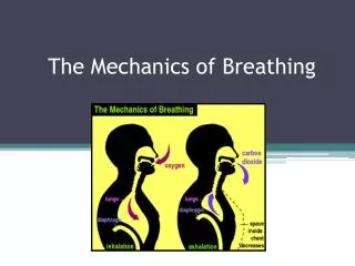
Remember that the process of inhalation brings O2 into the body whilst exhalation removes CO2. So, how does our breathing system enable us to do this. Well, inhaling and exhaling are brought about by certain changes in the position of our breathing system. Let us look again at the general structure of this system. Remember, the breathing system is found in the upper region of the body. This is known as the thorax.
Picture of the respiratory system Trachea Ribs Rib muscles Right Bronchus Diaphragm Right Lung Left Lung This system does not have a fixed shape. It has the ability to move, whilst remaining enclosed within the protection of the ribcage.
This means that the rib cage must also be able to change position. OBSERVATION Take your hands and place them flat on your chest just above your hips on each side of your body. Now breathe in and out very deeply. Whilst you do this, watch to see what happens to your hands. You should notice the following things…..
When you breathe in (inhale), your hands move up and outwards. When you breathe out (exhale), your hands move down and inwards. Inhaling Let’s see why…. When we inhale, our lungs fill with air. As they fill, they become enlarged. The ribs must then move upwards and outwards to make more room in the thorax. The overall effect of this is that our chest expands.
Your diaphragm is also involved in the inhalation process. It’s location beneath the lungs means that it separates the thorax from the abdomen. It is a sheet of muscle that spans the width of the body. Just before we inhale, it is found in a dome shape. As we inhale, it contracts and flattens. The result of this change in shape is a change in the volume of the thorax. Inhaling
As the volume of the thorax increases, the internal air pressure drops. This means that the air pressure outside the lungs is greater than the air pressure inside the lungs. High Low • Diaphragm flattens • High air pressure outside • Thorax volume increases • Low air pressure inside • Air pressure drops • Air diffuses into the lungs
If these changes occur when we breathe in, the opposite must happen when we breathe out. These changes can be summarised in the table below... Feature Inhaling Exhaling Flat Domed Diaphragm shape Up and out Down and in Ribs Contracted Relaxed Diaphragm muscle Contracted Relaxed Rib Muscle Inflated Deflated Lungs
Click on the ‘Air Drawn in’ buttons to explore the animation.
Click on the ‘Passage of air’ buttons to explore the animation.
“A Load of hot air!” The following activity will help you review your understanding of the structure of the breathing system. • Instructions: • There are 20 questions to answer • The number in brackets tells you how many letters in the word.
1 This is the toxic gas that is released when we breathe out? (6, 7) 2 When these contract and relax they move the rib-cage out and in? (3, 7) 3 The name for the minute air sacs that are covered with blood vessels? (7) 4 The area of the body where the lungs are found? (6) 5 A protective structure surrounding the lungs (3, 4) 6 The trachea branches into _________ (7), one going to each lung. 7 The circulatory system will take oxygen to the _______ (5) of the body. 8 Directly beneath the lungs is a sheet of muscle known as the ____________ . (9) “A Load of hot air!” Questions
9 The cavity through which we breathe and eat. (5) 10 The human breathing system contains two of these large spongy air bags. (5) 11 The other name for the wind-pipe? (7) 12 The name for the junction between the oesophagus and the wind-pipe? (7) 13 This is one of the dead-end sacs at the end of the bronchioles (8) 14 The left ________ (8) connects the left lung to the trachea. 15 The gas needed by the body to perform respiration? (6) 16 The diaphragm separates the breathing system from the __________.(7)
17 The area that connects the nose to the pharynx. (5, 6) 18 The main purpose of the breathing system is to generate _________. (6) 19 One of two openings of the breathing system located above the mouth. (7) 20 The name of the release of energy from food? (11)
Activity “Don’t hold your breath!” Pretending you are air! List down the answers to questions 19, 17, 13, 12, 11, 10 and 6. Now re-order the words to represent a trip through the breathing system, beginning outside the body. You must also try to fit the bonus word ‘bronchioles’ into your list.
Activity Where’s the air going? Use answers from questions 1, 7, 18, 15 and 20 to fill in the following explanation of the purpose of breathing oxygen into our bodies. When we breathe, we are doing so to fuel the process of ____________ , which is one of the characteristics of life. __________ is taken in and ________ __________ is removed in the process. The oxygen eventually leaves the breathing system and enters the circulatory system. This then transports the gas around the body to all the body ________ which can generate __________.
Activity Words that mean the same thing. Answer the following questions. Be careful, the spelling is essential! 1. What is the name of one tiny air sac? 2. Many tiny air sacs are known as ________? 3. What is the name of one branch of the trachea? 4. The name for the tubes that branch from the trachea are known as ___________?
Activity The lungs have to move! When we breathe in, our lungs fill with air. Identify two ways our breathing system creates more room in the thorax for these inflated lungs.